What Are Neuron-Derived Exosomes and Why Should You Care?
Understanding Neuron-Derived Exosomes
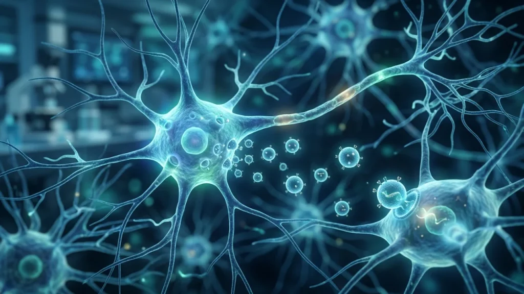
Imagine your brain’s billions of neurons need to send packages. They don’t use a postal service. They create their own tiny bubbles. These bubbles are called neuron-derived exosomes. They are incredibly small. You could line up thousands across the width of a single hair.
For a long time, scientists thought these bubbles were just garbage bags. They believed neurons used them to throw out unwanted material. That view has completely changed. Now we know these exosomes are vital communication tools. They carry important signals from one cell to another.
Think of a neuron like a factory. It constantly makes proteins and genetic material. It also produces these exosomes. The cell carefully loads each tiny bubble with specific cargo. This cargo can include: – MicroRNAs, which are instructions for other cells. – Proteins that can change how a receiving cell works. – Even pieces of damaged proteins that need removal.
The bubble itself is made of a lipid membrane. This is like a tiny cell membrane. It protects the precious cargo inside. It also has address labels on its surface. These labels help the exosome find the right target cell.
The release process is active, not passive. A neuron doesn’t just dump these vesicles. It guides them to its outer membrane. The bubble fuses with the cell wall and is released into the space between cells. This space is called the interstitial fluid. From there, exosomes can travel near or far.
Their journey is a key part of brain health. Neuron-derived exosomes can be picked up by neighboring cells. An astrocyte, a star-shaped support cell, might grab one. A microglia, the brain’s immune cell, might consume another. The cargo inside then changes what that cell does.
This system allows for precise messaging without direct wiring. Not every neuron is directly connected. Exosomes help them communicate anyway. They send signals that can tell another cell to grow, to calm down, or to prepare for stress.
Research shows their role is critical in disease too. In Alzheimer’s disease, neurons might send exosomes carrying toxic proteins. These could spread damage. In contrast, healthy exosomes might deliver helpful materials that protect cells.
Understanding this changes how we see the brain. It is not just a static network of wires. It is a dynamic, chattering community. Tiny lipid bubbles carry constant conversations between cells. This chatter maintains memory, learning, and overall brain function.
The term “cellular debris” is now outdated for these structures. Calling them debris is like calling a text message paper trash. The container might be disposable, but the information inside is valuable. Neuron-derived exosomes are the brain’s sophisticated text message system.
They are essential for everyday brain operations. Without this messaging, neural networks would fail to coordinate. Their study opens new windows into brain health and disease. By reading these messages, scientists hope to detect problems early. They could also create new treatments that use this natural delivery system.
This fundamental shift—from waste to messenger—is why you should care. It reveals a hidden layer of brain communication. This layer is crucial for everything your mind does.
Why These Tiny Vesicles Matter for Brain Health
Think of your brain as a bustling city. Neuron-derived exosomes are its postal and maintenance trucks. They do not just carry casual notes. They deliver vital supplies and urgent instructions that keep the city running smoothly every single day.
One key job is maintenance. Neurons constantly need new parts to stay healthy. Exosomes deliver these parts directly. They carry building blocks like proteins and lipids. They also bring genetic instructions in the form of RNA. This helps neighboring cells repair themselves. It also helps them adapt to new challenges.
For example, during learning, brain cells form new connections. This process requires specific materials. Exosomes shuttle these materials to the right place at the right time. They support the physical changes that store a memory. Without this delivery service, learning would be slower. Memories might not stick.
These vesicles also manage waste. Brain cells produce metabolic trash and damaged proteins. Some of this waste gets packaged into exosomes. The cell sends it out for disposal. Other cells, like microglia, can then clean it up. This is a crucial daily chore. It prevents toxic buildup inside neurons.
Another critical role is defense. The brain has its own immune signaling. When stress or inflammation occurs, neurons can release special exosomes. These nanoscale messengers carry alarm signals or protective compounds. They can tell other cells to raise their defenses. They can also deliver antioxidants to calm oxidative damage.
Consider a lack of sleep. It stresses brain cells. In response, they may alter the cargo of their exosomes. The vesicles might carry more stress-related proteins or protective microRNAs. This alerts the neural network to the problem. It is a form of cellular adaptation.
The health of this system affects your mental sharpness. Consistent, clean exosome signaling supports cognitive functions. – It aids focus by regulating neurotransmitter systems. – It supports mood by transporting factors that influence neuroplasticity. – It maintains energy by coordinating metabolic responses between cells.
When the exosome system works well, the brain is resilient. It can recover from daily stresses more effectively. It can maintain clearer communication pathways. This contributes directly to a sense of overall wellness and stable cognitive function.
Disruption in this process has clear consequences. If exosomes start carrying harmful cargo, problems begin. Toxic proteins may spread. Inflammatory signals can circulate. This is seen in age-related decline and neurodegenerative diseases. The daily maintenance fails. The city’s services break down.
Therefore, caring about neuron-derived exosomes means caring about your brain’s daily upkeep. Their function is a fundamental pillar of brain health. They are not just for rare events. They are active participants in your moment-to-moment mental state. Supporting the mechanisms that keep this system healthy could be a key to long-term cognitive vitality. This understanding naturally leads us to ask how we might protect or enhance this vital messaging system for better brain healthspan.
The Basic Structure of Neuron-Derived Exosomes
Think of a neuron-derived exosome as a tiny, specialized delivery truck. It is incredibly small. You could line up thousands of them across the width of a single human hair. Its design is not random. Every part has a job.
The most important part is its outer wall. This is called the lipid bilayer. “Lipid” means fat. “Bilayer” means two layers. This wall is like a flexible, protective bubble. It seals the exosome’s cargo safe inside. It also protects the cargo from the messy environment outside the cell.
This membrane wall is not plain. It is studded with important molecules. These molecules act like address labels and keys. – Some are used for targeting. They help the exosome find and dock with the correct recipient cell. – Others are for identification. They signal that this package came from a neuron.
Inside this protective bubble is the cargo. This is the exosome’s message. The cargo is a precise mix of molecules selected by the neuron. It is never just trash.
The main types of cargo include proteins. These can be tools for the receiving cell. Some proteins are enzymes that start chemical reactions. Others are signals that tell the cell to grow or change.
Another crucial cargo is RNA, especially microRNA. These are not blueprints for proteins. Instead, they are like instruction manuals or switches. They can turn specific genes in the receiving cell on or off. This changes what that cell does.
The exosome also carries other elements. It can have pieces of the cell’s skeleton inside. It even carries bits of the neuron’s outer membrane on its own surface. This gives the recipient cell a sample of the sender’s state.
The structure is built for purpose. The lipid wall merges easily with another cell’s membrane. This allows direct delivery. The addressing molecules ensure the message gets to the right place. The protected interior keeps fragile RNA intact during its journey.
This precise packaging is what makes neuron-derived exosomes so effective. They are not simple bags of leftovers. They are engineered communication devices. Their design allows them to travel through brain fluid. They can cross barriers and reach distant cells.
Understanding this structure explains their role in health and disease. A well-built exosome delivers helpful instructions. A damaged or misloaded one can deliver harmful cargo. The next section will explore how these tiny trucks are actually made and launched from the neuron.
How Neurons Create and Release Exosomes
Neurons do not randomly shed their parts. Creating an exosome is a careful, multi-step process. It happens inside the cell. Think of it as a specialized packaging assembly line. This line ensures only the right cargo gets loaded into the right container.
The journey starts in a compartment called the endosome. The cell’s membrane folds inward. It traps material from outside the cell or from its own surface. This forms an early endosome. It is like a sorting hub inside the cell.
The endosome’s membrane then buds inward. It creates tiny vesicles inside the larger compartment. This structure is now called a multivesicular body, or MVB. The MVB is a bubble filled with smaller bubbles. Those inner bubbles will become neuron-derived exosomes.
Cargo selection is critical at this stage. The cell actively chooses what goes into each tiny inner vesicle. Special protein complexes act as sorting machines. They recognize specific signal tags on molecules like RNA and proteins. These machines guide the chosen cargo into the forming vesicles.
This sorting decides the message’s content. A healthy neuron might pack growth signals and repair tools. A stressed neuron could pack distress signals or toxic proteins. The process is highly controlled but can be influenced by the cell’s condition.
Once loaded, the MVB has two possible fates. It can travel to the cell’s digestive center. There it merges with a structure called a lysosome. Its contents are broken down for recycling. This is the waste disposal path.
But for communication, the MVB takes a different route. It moves along the cell’s internal railway tracks. These tracks are made of proteins called microtubules. The MVB travels to the outer membrane of the neuron.
The final step is release. The MVB docks at the inner surface of the cell’s membrane. The membranes of the MVB and the cell fuse together. This fusion opens the MVB to the outside world. The inner vesicles are ejected into the extracellular space. They are now free neuron-derived exosomes, ready for delivery.
The entire process is energy-intensive. The neuron uses fuel to power it. This shows exosome release is not passive leakage. It is an active, deliberate form of communication.
Several factors can change exosome production. Increased neuronal activity often increases release. Cellular stress, like heat or lack of nutrients, can also trigger more release. Disease states can hijack the system, causing faulty packaging.
- Key steps in exosome creation:
- Formation of an early endosome as a sorting hub.
- Creation of intraluminal vesicles inside a multivesicular body.
- Active sorting and loading of specific molecular cargo.
- Transport of the loaded multivesicular body to the cell periphery.
- Fusion with the plasma membrane and release of exosomes.
This controlled production system is vital for brain function. It allows neurons to send precise signals to neighbors near and far. The next logical question is about their journey after release. How do these packages navigate the complex environment of the brain to find their target?
Key Differences Between Exosomes and Other Cell Parts
Neuron-derived exosomes are not the only packages a cell sends out. Cells release several types of tiny vesicles. It is crucial to tell them apart. Their origins and contents are different. This changes their function.
Think of a cell as a busy shipping department. It has two main ways to send out freight. The first way is like budding a package directly off the dock wall. The second way is like loading a shipping container inside the warehouse and then sending the whole container out.
Microvesicles use the first method. They pinch directly off from the cell’s outer membrane. This process is called budding. It happens at the plasma membrane itself. The cell’s membrane simply bulges outward. Then it seals shut at the base. This releases a vesicle into the space outside.
In contrast, neuron-derived exosomes use the second, more complex method. As detailed earlier, they form inside the cell. They are first created as tiny bubbles inside a larger container. This container is the multivesicular body. Only later does this whole container fuse with the cell’s wall. This releases the inner bubbles as exosomes. This two-step journey is their key signature.
This difference in origin leads to major differences in cargo. What gets packed inside is not random.
Because microvesicles bud directly from the surface, they carry lots of material from the cell’s outer membrane. Their cargo reflects the cell’s immediate surface state. It can include proteins that were just sitting on the membrane.
Exosome cargo is selected much earlier. It is chosen inside the cell’s sorting hubs. This allows for precise control. Neurons can pack specific signals into exosomes. These signals include: – MicroRNAs, which are instructions that can turn genes off in a target cell. – Protective or toxic proteins linked to brain health. – Bits of metabolic waste that need removal.
Size is another practical difference. Both are extremely small, measured in nanometers. But exosomes have a more uniform and smaller range. They are typically between 30 and 150 nanometers wide. A nanometer is one billionth of a meter.
Microvesicles are generally larger. They often range from 100 to 1000 nanometers. They are also more varied in size. Their shape can be less regular too.
Why does this distinction matter for brain science? The different creation paths mean these vesicles have different jobs.
Microvesicles often act in more local, immediate responses. They might help a cell quickly shed unwanted surface proteins. They can signal to very close neighbors about sudden stress.
Neuron-derived exosomes are built for targeted, long-distance communication. Their protected double-membrane structure makes them sturdy. They can travel farther in the brain’s fluid without breaking apart. Their pre-selected cargo is like a prepared message, not a quick note.
Researchers can separate them in the lab using advanced centrifuges. Exosomes are denser and sink at very high speeds. This lets scientists study their unique contents separately.
Confusing these two types leads to muddy data. Early studies often mixed them together. Modern research carefully isolates exosomes. This reveals their true role as sophisticated messengers, not just cellular debris.
Understanding this difference is key to grasping their potential. In brain diseases, both types of vesicles change. But exosome cargo might give earlier and more specific clues about neuronal health. Their precise origin makes them a cleaner window into the neuron’s inner world.
Knowing how exosomes differ from microvesicles sets the stage for their journey. Next, we will explore how these precise packages navigate the brain’s complex landscape to deliver their messages.
How Neuron-Derived Exosomes Work in the Healthy Brain
The Journey of Neuron-Derived Exosomes Between Cells
Neuron-derived exosomes begin their mission inside the cell. They form inside compartments called multivesicular bodies. Think of these as sorting stations. Here, the cell carefully packs molecules into the tiny vesicles. This cargo includes proteins, lipids, and genetic instructions like RNA. Once packed, the multivesicular body moves to the cell’s outer membrane. It fuses with this membrane. This action releases the exosomes into the space outside the neuron. This space is filled with fluid.
This release is not a random spill. It is a controlled export. Neurons can release more exosomes when they are active. Signaling from other cells can also trigger release. The exosomes are now free in the brain’s extracellular space. This is a crowded environment. They must navigate through a mesh of other cells and support structures.
Their journey is not a passive float. They move via diffusion in the brain’s fluid. They can also be guided by signals on their surface. These surface signals act like addresses or docking codes. They help the exosome find its target cell. The target could be another neuron nearby. It could also be a supporting glial cell, like an astrocyte or microglia, farther away.
The lipid membrane of the exosome is key for this trip. It protects the precious cargo from enzymes that would break it down. This double-layer shield ensures the message arrives intact. It allows for long-distance travel within the brain’s complex networks.
Delivery is the final critical step. The exosome must transfer its information to the target cell. Scientists have identified several ways this can happen. – The exosome can dock directly onto the target cell’s membrane. Surface proteins on both interact. This docking sends a signal directly into the target cell. – The target cell might engulf the entire exosome. It pulls the vesicle inside itself. Once inside, the exosome’s membrane degrades. This releases the cargo into the target cell’s interior. – The membranes of the exosome and the target cell can fuse. This fusion dumps the cargo directly into the target cell’s cytoplasm.
The method used depends on the cell types and the message. The result is always a change in the receiving cell. The delivered cargo can alter the cell’s behavior. For example, RNA from the exosome can be used by the target cell to make new proteins. These new proteins can change how the target cell functions or communicates further.
This process creates a continuous network of chatter. Neuron-derived exosomes allow neurons to talk to cells they are not directly wired to through synapses. They send metabolic signals. They share stress responses. They help coordinate immune activity in the brain by alerting glial cells.
In a healthy brain, this system is balanced and precise. It supports learning, memory formation, and overall brain maintenance. Exosomes help remove waste proteins from neurons. They also deliver nutrients and growth factors to areas in need.
The entire journey—from formation, to release, to transit, to delivery—shows these vesicles as active agents. They are not debris. They are essential communication packets. Their work connects different brain regions and cell types into one integrated system.
Understanding this precise journey in health gives us a baseline. We can now see what goes wrong when this system breaks down in disease. Faulty packaging, misdirected travel, or incorrect signals can disrupt vital brain communication.
What Neuron-Derived Exosomes Carry Inside Them
Neuron-derived exosomes carry a precise molecular toolkit. Think of them as tiny shipping containers. Each container holds a specific set of instructions and tools for the cell that receives it.
The cargo is not random cellular junk. It is carefully selected and packed. This selection happens inside the neuron’s multivesicular body before the exosome is released. The contents reflect the neuron’s current state and needs.
What fills these nanoscale messengers? The cargo falls into three main groups.
First are nucleic acids, mainly RNA. This is genetic instruction material. The most common type is microRNA, or miRNA. These are short strands of RNA. They do not code for proteins themselves. Instead, they act as managers. They can silence specific genes in the target cell. One neuron-derived exosome might carry miRNA that tells a glial cell to calm inflammation. Another might carry RNA that helps a neighboring neuron strengthen its connections.
Second are proteins. These are the workhorse molecules of the cell. Exosomes carry hundreds of different protein types. Some are common to all exosomes. These include proteins used for structure and fusion. Others are unique signals from the neuron.
Key protein categories include: – Receptors and ligands. These sit on the exosome’s surface. They act like keys to unlock specific target cells. – Enzymes. These drive chemical reactions inside the receiving cell. – Growth factors. These encourage cells to grow, survive, or change. – Prion proteins. In their normal form, these proteins help with neuron signaling and protection.
Third are lipids. The exosome’s membrane itself is made of a lipid bilayer. The specific lipids present are not just a shell. They aid in stability, targeting, and fusion with the target cell’s membrane.
This cargo mix is dynamic. A neuron under stress will pack different cargo than a neuron involved in forming a memory. For example, during learning, exosomes may carry more proteins related to synaptic plasticity. During metabolic stress, they might carry more signals to request energy support.
The combination is what makes the message. An exosome carrying a certain miRNA plus a specific growth factor creates a compound instruction. It is like sending a written note along with a physical key. The target cell reads both and responds accordingly.
This precise packaging ensures that brain communication is accurate. A message meant for an astrocyte should not accidentally activate a microglial immune response. The surface markers and internal cargo work together for specificity.
The contents also allow for long-term changes. Proteins can have an immediate effect on the receiving cell. RNA can alter that cell’s gene expression for hours or days. This means one exosome can have a lasting impact.
Understanding this cargo list is crucial. In a healthy brain, the right tools are sent to the right places at the right time. This supports all brain functions. It keeps neural networks tuned and resilient.
We now know what these messengers carry in health. This gives us a clear inventory. We can spot when the cargo list goes wrong in disease states, which often involves toxic proteins or misleading signals.
How Exosomes Help Brain Cells Talk to Each Other
Neuron-derived exosomes are the brain’s private courier service. They carry vital messages from one cell to another. This system is fast and precise. It works over both short and long distances.
Think of a neuron sending a signal. It fires an electrical pulse down its axon. This is one type of communication. But it is like shouting across a room. Exosome communication is different. It is like handing someone a sealed envelope with detailed instructions inside.
The process starts with a need. A neuron might need help from a nearby astrocyte. Astrocytes are support cells. They provide nutrients and manage waste. The neuron creates an exosome with a specific request. The exosome pinches off into the space between cells.
This space is called the interstitial fluid. It is the brain’s environment. The exosome navigates this fluid. It does not travel randomly. Proteins on its surface act like an address label. These labels guide it to the correct cell type.
For example, an exosome meant for an astrocyte will have markers that astrocytes recognize. A microglial cell would ignore this package. This targeting ensures messages go to the right place.
Delivery is the next step. The exosome reaches its target cell. It docks onto the cell’s membrane. The membranes then fuse together. The exosome’s cargo spills directly into the receiving cell’s interior.
This direct delivery is key. It protects fragile molecules like RNA during the journey. Nothing gets lost or degraded in transit. The message arrives intact and ready to use.
The effect on the receiving cell can be immediate. Delivered proteins can start working at once. They might trigger a new signal inside the cell. They could change how the cell uses energy.
Other effects take longer. RNA cargo is a set of blueprints. The receiving cell reads these blueprints. It then makes new proteins based on the instructions. This changes the cell’s function for hours or even days.
Let’s look at a concrete example in memory formation. – Neurons are active when we learn something new. – These neurons release exosomes packed with specific miRNAs. – These exosomes travel to neighboring neurons. – The miRNAs help strengthen the connections between those neurons. – This strengthening is the physical basis of a memory.
Exosomes also help with routine maintenance. Neurons constantly need support. They send out requests for nutrients. They also need to remove waste proteins.
Astrocytes often fulfill these requests. An exosome from a neuron can signal an astrocyte to deliver more lactate, which is a fuel source. Another exosome might carry damaged proteins for the astrocyte to break down and recycle.
This cleanup is vital for brain health. It prevents toxic junk from building up inside neurons.
Communication is not one-way. Other brain cells send exosomes back to neurons. Astrocytes and microglia release their own vesicles. These messages can tell a neuron to grow, to calm down, or to prepare for activity.
This creates a constant, buzzing conversation. Millions of these tiny messages are exchanged every second in your brain. They coordinate everything you think and feel.
The system is remarkably robust. If one pathway is blocked, cells can sometimes use another. They might send a different type of exosome or adjust the cargo.
This flexibility keeps the brain resilient. It allows neural networks to adapt and rewire themselves based on experience.
In summary, neuron-derived exosomes are fundamental to brain talk. They enable targeted, complex, and lasting communication between all brain cells. This dialogue maintains the health and function of the entire system. Understanding this language is the first step to seeing what happens when it breaks down in disease.
The Role of Neuron-Derived Exosomes in Memory and Learning
Memory formation is not a static event. It is a physical change in your brain’s wiring. These changes happen at connections called synapses. A synapse is where one neuron talks to another. For learning to stick, these synapses must get stronger or weaker. This ability is called synaptic plasticity.
Neuron-derived exosomes are essential tools for this job. They help remodel the synapse from both sides. Think of a synapse as a busy dock. The sending neuron has a presynaptic terminal. The receiving neuron has a postsynaptic spine. Both sides use exosomes to ship crucial supplies.
The sending neuron packages specific cargo into exosomes. This cargo can include: – Building blocks for neurotransmitters, the chemical signals. – Proteins that help release these signals faster. – Signals that tell the receiving cell to get ready.
These exosomes travel the short distance to the receiving neuron’s spine. They deliver their cargo directly to the site. This strengthens the communication line. It makes the signal clearer and faster next time.
The receiving neuron also sends exosomes back. Its cargo might include: – Receptor parts to catch more neurotransmitter. – Enzymes that stabilize the spine structure. – Feedback signals to fine-tune the sender’s activity.
This two-way exchange solidifies the connection. It is like both sides agreeing to upgrade their equipment. This upgrade is the physical basis of a memory.
A key process is called Long-Term Potentiation, or LTP. LTP is a long-lasting increase in signal strength. It is a major cellular model for memory. Research shows exosomes are active during LTP. Blocking their release can impair this strengthening.
Exosomes carry molecules that trigger growth. They deliver proteins like BDNF, or brain-derived neurotrophic factor. BDNF acts as a fertilizer for synapses. It encourages spines to grow larger and more stable. A larger spine can hold stronger connections.
These vesicles also remove unwanted material. To make room for new memories, old proteins must be cleared. Exosomes carry away used or damaged molecules from the synapse. This cleanup prevents clutter and allows for new growth.
The timing of this traffic is precise. Learning events cause a surge in exosome release. Neurons dispatch these messengers exactly when needed. This ensures changes happen at the right synapse at the right moment.
Different types of learning use different exosome cargos. Spatial learning might involve one set of proteins. Fear conditioning might involve another set. The cargo is tailored to the task, creating a specific molecular signature for each memory.
This system allows for efficiency and specificity. Instead of flooding the whole cell with signals, neurons send targeted packages. This precision prevents cross-talk between unrelated memories. It keeps your knowledge of a friend’s face separate from your memory of a recipe.
The role of neuron-derived exosomes extends beyond initial formation. They are also involved in memory consolidation. This is when a fragile short-term memory becomes a durable long-term one.
During sleep, especially deep sleep, this exosome traffic may increase. The brain replays the day’s activities. Exosomes help solidify the synaptic changes made during wakefulness. They integrate new information into existing networks.
Without this vesicle-based system, synaptic plasticity would be slow and messy. Neurons would struggle to coordinate precise changes across tiny gaps. Exosomes provide a direct, fast, and information-rich delivery service.
They enable the brain’s constant rewiring. Every new fact you learn, every skill you practice, involves these nanoscale messengers. They are the couriers of change, turning fleeting experiences into lasting neural structures.
Understanding this link explains why disrupting exosome function can harm cognition. If the delivery system fails, synapses cannot adapt properly. Memories become hard to form or easy to lose.
The healthy brain relies on this flawless molecular logistics network. Neuron-derived exosomes ensure that our life experiences leave a permanent, physical trace in the intricate architecture of our minds.
Exosomes and Brain Repair Mechanisms
The brain has a remarkable capacity to mend itself after minor injuries. Neuron-derived exosomes are central to this internal repair process. They do not just carry messages for learning. They also deliver emergency supplies to damaged sites.
Think of a tiny scratch on a neuron’s surface. This can happen from normal stress or inflammation. The cell quickly responds. It releases special exosomes loaded with repair tools. These vesicles travel to the exact spot that needs help.
What is inside these repair packages? The cargo is carefully selected. It often includes growth factors. These are proteins that tell cells to grow and heal. They also contain lipids to patch damaged cell membranes. Some carry instructions in the form of RNA to help the neuron rebuild its structures.
This targeted delivery is far more efficient than releasing signals into the wide space between cells. It prevents wasted effort. It also avoids accidentally stimulating nearby healthy neurons. The system is precise and local.
Exosomes help clean up brain debris too. During damage or disease, toxic proteins can accumulate. These harmful clumps can kill neurons. Exosomes can bind to this debris. They help move it to cells that break it down for removal. This cleanup keeps the neural environment healthy.
The vesicles also aid in reducing inflammation. Brain inflammation is a common response to injury. But too much inflammation causes more harm. Neuron-derived exosomes can carry anti-inflammatory signals. They tell immune cells in the brain to calm down. This limits collateral damage to healthy tissue.
Here is a simplified sequence of how they assist in repair: – A neuron experiences stress or detects damage in a connected cell. – It packages specific repair molecules into exosomes. – The exosomes are released and navigate to the target site. – They fuse with the recipient cell or are taken inside. – The cargo is unloaded and begins its healing work.
This process is continuous at a low level. It maintains the brain’s vast network every day. When injury occurs, the activity of these exosomes can increase dramatically. They become part of the brain’s first-response team.
Their role is crucial for recovery from events like a minor stroke or concussion. In such cases, communication lines are cut. Exosomes help re-establish connections. They support surviving neurons as they form new pathways around the damaged area.
Research shows these vesicles can even encourage neurogenesis in some brain regions. Neurogenesis is the birth of new neurons. Exosomes from healthy neurons can carry signals that prompt stem cells to divide. They guide the development of new, functional nerve cells.
This repair function relies on the same core features that aid memory: precise targeting and rich information transfer. The difference lies in the cargo and the goal. For memory, the cargo changes synaptic strength. For repair, it delivers survival and reconstruction signals.
Without this exosome-mediated system, the brain would be far more fragile. Small everyday wear and tear would accumulate. The system provides a dynamic maintenance program. It constantly patches, cleans, and reinforces our neural circuitry.
Understanding this opens new perspectives on brain health. A robust exosome system may contribute to resilience against age-related decline. It represents a natural defense mechanism working at the nanoscale.
The health of a neuron influences the quality of its exosomes. A stressed or sick neuron might send out confusing or harmful signals. This highlights the interconnected nature of brain cell networks. The failure of one cell’s communication can impact many others.
Ultimately, neuron-derived exosomes are multitaskers. They are essential for adapting the brain through learning. They are equally vital for protecting and restoring it from damage. This dual role makes them indispensable for lifelong brain function. Their continuous activity in the background is a key reason our brains can last a lifetime, adapting and recovering from countless small challenges. This built-in repair capacity paves the way to understanding what happens when these systems falter, as seen in various neurological diseases.
Neuron-Derived Exosomes in Brain Diseases
How Exosomes Change in Alzheimer’s Disease
In Alzheimer’s disease, the communication system using neuron-derived exosomes goes wrong. Neurons under stress start sending out harmful signals. These signals can speed up the disease process.
The cargo inside these vesicles changes dramatically. Two toxic proteins linked to Alzheimer’s are often found packed inside. These proteins are amyloid-beta and tau.
Healthy exosomes might help clear these proteins away. In Alzheimer’s, this process seems to break down. The exosomes instead may spread the proteins to other cells.
Think of it like a postal service delivering poison instead of food. The receiving cell gets damaged. This can cause a chain reaction of damage across brain networks.
The exosome membrane itself can also change. Its surface markers might become altered. This could affect where the vesicle ends up going in the brain.
Scientists can see these changes in real patients. They study exosomes from blood or spinal fluid. The exosomes from people with Alzheimer’s look different.
Their molecular cargo acts like a fingerprint of disease. Researchers look for specific signatures. This includes: – High levels of toxic phospho-tau protein. – Low levels of protective synaptic proteins. – Distinctive microRNA patterns linked to inflammation.
This is not just a passive reflection of damage. Evidence suggests these altered exosomes actively harm synapses. They can disrupt the very connections needed for memory.
The inflammatory signals they carry create a hostile environment. Support cells called microglia get activated. This leads to chronic inflammation that harms neurons.
It becomes a vicious cycle. Diseased neurons send bad exosomes. These exosomes cause more damage and stress. Then more stressed neurons send more bad exosomes.
This cycle helps explain how Alzheimer’s progresses slowly. The exosome system might fail gradually over years. Small communication errors accumulate into major network failure.
Diagnostically, these neuron-derived exosomes offer a huge advantage. They provide a direct window into brain processes from a simple blood draw. They are like tiny messengers reporting on the health of their neuron of origin.
Current research focuses on two main goals. The first is early detection. Finding these altered exosomes could flag Alzheimer’s long before symptoms appear.
The second goal is treatment. Could we engineer healthy exosomes to deliver helpful drugs? Could we block the release of the harmful ones?
Understanding this faulty communication is key to stopping the disease. It shifts the view from just plaques and tangles to a dynamic signaling problem. The brain’s own messaging system becomes part of the pathology.
This reveals a critical insight for brain health. Maintaining clean cellular communication is as important as removing waste. When the messages themselves become corrupted, the entire system falters.
The story in Alzheimer’s shows the dark side of this powerful biological tool. The same system that maintains and adapts the healthy brain can, when corrupted, accelerate its decline. This duality makes targeting exosome pathways a promising but complex frontier for therapy, leading naturally to questions about their role in other neurological conditions.
The Impact of Neuron-Derived Exosomes on Parkinson’s
In Parkinson’s disease, a specific protein called alpha-synuclein clumps together inside neurons. These clumps are a hallmark of the disease. They damage and eventually kill brain cells. For years, a major puzzle was how the disease seems to spread through the brain. The pathology starts in one area and then moves to others. Neuron-derived exosomes provide a compelling answer to this puzzle.
Think of these exosomes as tiny cargo ships. They can pick up abnormal alpha-synuclein from a sick neuron. The exosomes then carry this harmful cargo to neighboring healthy neurons. They deliver the misfolded protein like a toxic package. The receiving cell then often starts to form its own clumps. This process repeats, creating a chain reaction.
This is not just theory. Laboratory studies show it clearly. Researchers can collect exosomes from the fluid of Parkinson’s patients. They then add these exosomes to healthy neurons in a dish. The healthy cells begin to show signs of disease. They develop alpha-synuclein clumps and start to malfunction. This demonstrates the direct transmitting role of these vesicles.
The content of these neuron-derived exosomes tells a detailed story. They carry more than just alpha-synuclein. Their molecular cargo changes in Parkinson’s. Scientists find specific patterns inside them.
- They contain elevated levels of the pathological alpha-synuclein.
- They often carry markers of cellular stress and inflammation.
- They may have reduced levels of protective factors.
- Their lipid membrane composition can be altered.
This altered cargo does two harmful things. First, it spreads the core pathology. Second, it sends misleading signals to other cells. It can trigger inflammation from immune cells in the brain called microglia. This creates a hostile environment for all neurons.
The impact on brain networks is severe. Parkinson’s primarily affects movement control. This control depends on a circuit called the basal ganglia. The spread of toxic exosomes can disrupt this critical circuit. Communication between neurons breaks down. Signals for smooth movement get lost or corrupted.
Diagnostically, this offers new hope. Detecting pathogenic alpha-synuclein in blood-based neuron-derived exosomes could lead to earlier diagnosis. Today, Parkinson’s is often diagnosed only after significant cell loss has occurred. A test based on these vesicles could identify the process years earlier.
Therapeutic strategies are now targeting this system. The goal is to intercept the bad messengers. Several approaches are being explored in preclinical research.
One approach is to develop antibodies that grab the harmful exosomes. These antibodies could tag them for destruction before they deliver their cargo. Another idea is to engineer decoy receptors. These decoys would catch the exosomes like a net, preventing them from reaching healthy cells.
A different strategy aims at the source. Could we stop sick neurons from loading toxic cargo into exosomes? Or could we encourage them to release only helpful vesicles? This requires precise molecular tools to edit cellular export programs.
Understanding this exosome-mediated spread changes our view of Parkinson’s. It is not just a disease of isolated cells dying one by one. It is a disease of faulty communication where cells inadvertently harm their neighbors. The messaging system becomes a weapon.
This insight connects back to brain health broadly. It reinforces that neural communication must be clean and accurate. Corrupted messages have physical consequences. They can literally reshape the brain’s architecture over time.
The story in Parkinson’s powerfully mirrors that in Alzheimer’s, yet with its own unique protein culprit. It confirms that neuron-derived exosomes are a common pathway in neurodegeneration. This shared mechanism opens doors for broader treatments that might protect neural networks across different diseases by safeguarding cellular communication itself.
Exosomes and Multiple Sclerosis Inflammation
In Multiple Sclerosis, the body’s own immune system attacks the insulation around nerve fibers. This insulation is called myelin. Neuron-derived exosomes play a key role in this attack. They do not carry toxic proteins like in Parkinson’s. Instead, they carry inflammatory signals.
Think of a neuron under stress. It might be damaged by early inflammation. This neuron then releases special exosomes. These vesicles are loaded with specific cargo. This cargo can include: – Tiny RNA molecules that instruct immune cells to become more aggressive. – Proteins on their surface that act as “find me” signals for immune cells. – Bits of myelin debris that further provoke the immune response.
These exosomes travel from the stressed neuron to immune cells in the brain. The primary immune cells involved are called microglia and astrocytes. The exosomes deliver their inflammatory instructions. This turns a local problem into a wider alarm.
The immune cells become activated. They start releasing their own damaging chemicals. They also recruit more immune cells from the bloodstream. This creates a cycle of inflammation. The exosomes essentially fan the flames of the attack.
Research shows this is a two-way street. Activated immune cells also release their own exosomes. These immune cell exosomes can then travel back to neurons. They carry signals that can harm the neuron directly or make it more vulnerable. The dialogue becomes destructive.
This process helps explain the spread of MS lesions in the brain. An inflammatory lesion might start in one area. Exosomes can then carry the inflammatory message to nearby, yet unaffected, regions. They prepare the ground for new lesions to form. The disease progresses through this corrupted communication.
Scientists are measuring these exosomes in the spinal fluid of MS patients. The cargo inside these vesicles reflects the disease’s activity. More inflammatory cargo often means more active disease. This makes exosomes potential biomarkers. They could help doctors track the illness without invasive biopsies.
Therapeutic strategies are emerging from this knowledge. The goal is to intercept the inflammatory exosomes. One idea is to use molecules that block their release from neurons. Another approach aims to neutralize them after they are released.
Some experimental treatments try to clean up the exosome cargo. They aim to stop neurons from loading inflammatory signals into the vesicles in the first place. This is a form of molecular editing inside the cell. It requires great precision.
The story in MS reinforces a broader principle. Neuron-derived exosomes are not passive debris. They are active participants in disease. In Parkinson’s, they spread misfolded proteins. In Alzheimer’s, they can spread tau tangles. In MS, they spread inflammatory commands.
This shared pathway offers hope. Understanding how neurons use these vesicles to talk to immune cells gives us new targets. We might one day treat MS by correcting the messages themselves. We could tell neurons to send calming signals instead of alarms.
Protecting neural networks requires clean communication on all fronts. It requires accurate signals between neurons themselves. It also requires peaceful signals between neurons and the immune system. When this dialogue breaks down, disease follows.
The study of exosomes in MS provides a clear example of this breakdown. It shows how a neuron’s cry for help can be misinterpreted by the body’s defenses. This misinterpretation leads to a sustained attack on the very system it aims to protect.
This insight connects brain health directly to immune health. The brain is not an isolated fortress. It is in constant, delicate conversation with the body’s defense forces. Exosomes are critical messengers in that conversation.
Future therapies may look very different. They might not just suppress the immune system broadly. They could instead retrain the communication system. The focus shifts from stopping soldiers to fixing the corrupted orders they receive.
The role of neuron-derived exosomes in MS completes a troubling picture across brain diseases. Whether the cargo is a toxic protein or an inflammatory signal, the outcome is similar. The messaging system becomes a weapon against the brain itself. Safeguarding this system is therefore a universal goal for neurological health.
Why Faulty Exosomes Can Spread Brain Damage
Neuron-derived exosomes can turn from messengers into carriers of harm. This happens when their cargo becomes dangerous. A sick neuron might pack toxic proteins into these vesicles. It then sends them out into the brain’s extracellular space.
Think of it like sending a letter contaminated with a poison. The letter itself is not evil. But its contents are deadly. Neighboring cells receive this toxic mail. They open it and are harmed.
This process helps explain how damage spreads in diseases like Alzheimer’s. Healthy neurons connect in a vast, delicate network. One diseased neuron can start a chain reaction. It releases exosomes filled with sticky beta-amyloid or tangled tau proteins.
Receiving cells import these toxic proteins. Their own internal machinery then gets clogged. They too become sick. Soon, they also begin sending out bad exosomes.
The damage fans out across the neural circuit. This exosome-based spread is efficient and targeted. It follows the brain’s own communication highways.
The problem is not just toxic proteins. Faulty signals can also spread. In conditions like Parkinson’s, a protein called alpha-synuclein can misfold. Exosomes carry these misfolded forms to new cells.
The new cell then copies the misfold. Its own alpha-synuclein proteins tangle up. This is similar to a prion disease. The exosome delivers the template for corruption.
Chronic inflammation also uses this system. Stressed neurons can send exosomes with inflammatory signals. These signals activate the brain’s immune cells, called microglia.
The microglia become overactive. They start releasing harmful chemicals. This creates a toxic environment for neurons. More neurons get stressed and send more inflammatory exosomes.
A vicious cycle begins. The exosomes keep the inflammatory fire burning.
- They carry “activate” orders to immune cells.
- They distribute toxins to healthy neurons.
- They spread protein misfolding patterns.
This turns a local problem into a widespread disease. The brain’s elegant messaging system is hijacked.
The lipid membrane of the exosome makes this possible. It protects the cargo during transit. The cargo does not degrade quickly. It also helps the vesicle fuse precisely with target cells.
Delivery is guaranteed. This is perfect for normal signaling. It becomes disastrous for spreading damage.
Researchers see this in lab models. Blocking exosome release can slow disease progression. It does not stop the initial sick neuron. But it can limit the contagion.
This points to a new therapeutic idea. We might not always cure the first sick cell. Yet we can protect its neighbors. We can intercept the bad messages.
The goal is to stop the chain letter of damage. Treatments could filter out harmful exosomes. Other treatments could empty them of their toxic cargo before delivery.
Some approaches aim to change the message itself. Could we engineer exosomes to carry protective drugs? Could we make them send repair signals instead?
Understanding this spread is crucial. It shows that brain diseases are often communicative disorders. The units of communication are these tiny vesicles.
Faulty exosomes do more than carry debris. They actively participate in the disease process. They are tools for damage propagation.
This shifts our view of neurological decline. It is not always a simple wear and tear. It can be an active, cell-to-cell transmission of failure.
The next frontier is learning to control this system. We must learn to correct the mail before it delivers its poison. Safeguarding neural communication requires monitoring these nanoscale messengers constantly. Their cargo determines the health of the entire network.
Tracking Disease Through Neuron-Derived Exosomes
Neuron-derived exosomes carry molecular snapshots of the cells that created them. This makes them powerful tools for tracking brain health. Think of them as tiny diagnostic bottles thrown into the bloodstream. For the first time, we can get a direct look at living brain chemistry without a single incision.
Doctors usually rely on scans or spinal taps. These methods show structure or general inflammation. They rarely reveal specific cellular changes happening right now. Neuron-derived exosomes offer that precise window. Their cargo reflects the real-time state of their neuron of origin.
What exactly can we find inside these vesicles? The list is revealing. – Misfolded proteins like tau or alpha-synuclein signal Alzheimer’s or Parkinson’s. – Specific microRNA fragments can indicate stress, inflammation, or repair processes. – Faulty metabolic enzymes may point to energy failures in neurons. – Pieces of damaged mitochondrial DNA can show cellular distress.
The key is that these molecules are packaged and protected. They travel from the cerebrospinal fluid into the blood. Scientists have developed methods to capture these specific vesicles from a simple blood sample. They use antibodies that latch onto unique surface markers. This isolates neuron-derived exosomes from all others.
Research shows clear patterns. People with Alzheimer’s disease have exosomes packed with phosphorylated tau. Their levels rise years before clear symptoms appear. This is a major breakthrough. It moves diagnosis from observing symptoms to detecting hidden biology.
The potential for early detection is immense. Catching a disease process early changes everything. Interventions can start before widespread damage occurs. This could mean lifestyle changes, existing drugs, or future therapies applied at the optimal time.
Tracking is not just for diagnosis. It is for monitoring. A doctor could draw blood every few months. They could check if a new drug is reducing toxic cargo in exosomes. This gives a fast measure of treatment effectiveness. Today, assessing brain treatments can take years of clinical observation.
Consider this concrete example. Two patients start the same therapy. Their memory tests show similar slow decline after one year. However, their exosome profiles tell a different story. One patient shows a 60% drop in amyloid-beta peptides in neuron-derived exosomes. The other shows no change. The therapy is likely helping the first patient at a cellular level, even if symptoms lag.
This technology also helps distinguish between diseases. Depression, chronic fatigue, and early dementia can have overlapping symptoms. Their exosome signatures are different. A precise molecular profile guides correct treatment from the start.
The path forward involves creating standard panels. Labs will test for a suite of markers from a single sample. A person’s results would create a personal brain health baseline. Future deviations from this baseline would trigger alerts.
Challenges remain. Techniques need refinement for widespread clinical use. We must fully understand normal variation between healthy individuals. Yet the direction is set. The era of liquid biopsies for the brain has begun.
Neuron-derived exosomes are more than messengers of disease. They have become our informants. By learning to read their cargo, we gain an unprecedented advantage. We can see the hidden progression of brain disorders and measure our success in stopping them. This turns a mechanism of disease into a powerful tool for health. The next step is using this information not just to track illness, but to predict and prevent it proactively.
Using Neuron-Derived Exosomes for Medical Advances
Exosomes as Tools for Early Brain Disease Detection
Imagine finding a sealed message from a brain cell in a simple blood test. This is now possible. Neuron-derived exosomes offer a direct window into the living brain. They do this without surgery or complex scans. Doctors can detect trouble long before symptoms appear.
The process starts with a standard blood draw. The blood contains exosomes from all over the body. The key step is isolation. Scientists use antibodies to fish out only the neuron-derived exosomes. These antibodies latch onto unique proteins on the vesicle’s surface. These proteins act like a return address, confirming the exosome came from a neuron.
Once isolated, researchers open these tiny packages. They analyze the cargo inside. This cargo includes proteins, bits of genetic code called RNA, and other molecules. In a healthy brain, this cargo has a normal pattern. In a developing disease, the pattern changes dramatically.
For example, in Alzheimer’s disease, harmful proteins accumulate in the brain years before memory fades. Neuron-derived exosomes often carry traces of these proteins. They may contain abnormal forms of tau or amyloid-beta. Finding these markers in blood exosomes signals a problem is brewing. It is like smoke appearing long before the visible fire.
This method is far more sensitive than checking for these proteins floating freely in blood. The exosome protects its cargo from degradation. It delivers a concentrated, cell-specific signal. This allows for detection at very early stages.
The advantages of this approach are clear. – It is minimally invasive. A blood test is simple and low-risk. – It can be repeated often. This allows doctors to monitor changes over time. – It provides specific cellular information. The data comes directly from neurons, not other cell types.
Early detection changes everything for many brain conditions. In Parkinson’s disease, movement symptoms begin after significant neuron loss. Exosome tests might reveal biochemical shifts a decade earlier. This creates a critical window for intervention. Therapies could start when they are most likely to protect the brain.
The same logic applies to brain tumors and neuroinflammatory diseases. Cancer cells shed exosomes with distinct signatures. These signatures can reveal tumor type and even genetic mutations. Inflammation in the brain alters exosome cargo, signaling an immune system attack on neural tissue.
Of course, this is not a single test for all diseases. Researchers are building detailed libraries. They match exosome cargo profiles to specific disorders. A diagnostic panel might check for fifty markers at once. This creates a fingerprint for health and disease.
Validation is an ongoing task. Scientists must confirm that these exosome signals consistently appear before clinical diagnosis. They must define normal ranges across ages and populations. The work is complex but the potential is immense.
This technology moves medicine from reaction to prevention. We are no longer waiting for the brain to fail. Instead, we intercept its distress signals mailed out in microscopic vesicles. The path from a blood vial to a brain health report is now being paved by these extraordinary messengers.
How Scientists Study Neuron-Derived Exosomes in Labs
Scientists cannot study neuron-derived exosomes directly from brain tissue in living people. Instead, they get them from fluids like blood or spinal fluid. This is a major technical challenge. These fluids contain trillions of vesicles from many cell types. Finding the tiny fraction that comes only from neurons is like finding a specific needle in a stack of other needles.
The first step is isolation. Researchers use several methods to separate exosomes from other particles.
- Ultracentrifugation is a classic technique. Samples are spun at incredibly high speeds. These forces push heavier particles to the bottom first. Lighter exosomes gather later. This method is common but can be harsh.
- Size-based chromatography acts like a microscopic filter. The fluid flows through columns with tiny pores. Larger particles get trapped. Smaller molecules pass through. Exosomes of a specific size are collected in the middle fraction.
- Immunoaffinity capture is the most precise tool for finding neuron-derived exosomes. It uses antibodies. These are proteins that stick to one specific target. Scientists use antibodies that latch onto markers found only on the surface of neuron-derived vesicles. This pulls them directly from the mix.
After isolation, scientists must confirm what they have. They check the harvest using multiple tests.
One common test is electron microscopy. It takes pictures of the particles. Researchers can see the classic cup-shaped structure of exosomes. Another test measures size. Instruments called nanoparticle trackers count particles and measure their diameter. True exosomes are typically between 30 and 150 nanometers wide.
But the real treasure is inside the vesicle. To access the cargo, scientists break open the lipid membrane. They use detergents or sound waves to do this gently. The released contents are then analyzed.
Protein analysis is key. Techniques like Western blotting check for known neuronal proteins. These include synaptophysin or L1CAM. Finding these proves the exosomes came from neurons. More advanced mass spectrometry can list hundreds of proteins at once. This creates a cargo profile.
Genetic material inside is also studied. RNA is extracted and sequenced. This shows which genetic instructions the neuron was sending out. A neuron under stress may send different RNA than a healthy one.
All this data needs context. Lab studies often compare exosomes from different groups. For example, researchers might analyze samples from fifty healthy people and fifty with Alzheimer’s disease. They look for consistent differences in protein or RNA levels.
These differences become potential biomarkers. A biomarker is a measurable signal of a condition. Discovering one requires repeated verification across many samples.
Control experiments are critical. Scientists must prove their isolation method worked. They test for contaminants like non-neuronal cell debris. They also ensure their detection methods are sensitive enough to see small changes.
The entire process is like forensic science for cellular messages. Researchers collect microscopic evidence from body fluids. They authenticate its source. They carefully unpack its contents. Finally, they decode the meaning behind the molecular signals.
This lab work builds the foundation for every medical application. Without these precise techniques, we could not identify the diagnostic fingerprints mentioned earlier. The next frontier is streamlining these methods for clinical use, turning complex lab protocols into fast, reliable tests for doctors’ offices.
The path from a research lab to a hospital clinic is long but clear. Each careful experiment brings us closer to using these sophisticated nanoscale messengers in everyday medicine.
Engineering Exosomes for Targeted Brain Therapies
Scientists are now building custom exosomes in the lab. They start with natural neuron-derived exosomes. Then they change them to become precise medical tools. This field is called exosome engineering. The goal is to create targeted brain therapies.
Think of a natural exosome as a delivery truck. It has a natural address on it. This address sends it to certain brain cells. Engineers can change this address. They can put a new GPS destination on the truck’s surface. This new GPS is a special protein or antibody.
Why is this so important? The brain has a strict barrier. This is called the blood-brain barrier. It blocks most drugs from entering. Large molecules and conventional drugs cannot cross it. This is a major problem for treating brain diseases.
Neuron-derived exosomes offer a unique solution. They are natural brain messengers. Their lipid membrane is similar to brain cell membranes. This lets them cross the protective barrier. They can carry cargo directly into neurons.
So, what cargo can we load into them? Engineers can pack many things inside. – Small drug molecules for diseases like Alzheimer’s. – Therapeutic RNA to fix faulty genetic instructions. – Protective proteins to support sick neurons. – Enzymes to break down toxic clumps in brain cells.
The loading process is a key engineering step. Scientists use several methods. One method is electroporation. A quick electric pulse opens tiny holes in the exosome’s membrane. The drug slips inside. Then the holes seal shut. Another method incubates exosomes with the cargo under specific conditions. The cargo naturally passes through the membrane.
Surface modification is equally critical. An engineered exosome needs to find the right cell. Researchers can add targeting molecules to its outer shell. These molecules act like homing signals. They bind only to receptors on diseased neurons. This makes treatment precise and reduces side effects.
For example, a team might design an exosome for a brain tumor. They load it with a cancer-killing drug. Then they add a peptide that binds only to tumor cells. The exosome crosses the blood-brain barrier. It seeks out the tumor. It delivers its lethal payload directly to the cancer.
The potential is vast for neurodegenerative diseases. In conditions like Parkinson’s, specific neurons die. Engineered exosomes could deliver growth factors to those cells. They could tell them to survive and function better. They could also carry tools to remove harmful proteins like alpha-synuclein.
This work moves beyond just observation. We are no longer just reading the messages neurons send. We are now writing our own instructions. We are creating custom nanoscale messengers with clear medical orders.
The path from engineering to therapy requires more testing. Safety studies must confirm these designed vesicles do no harm. Large-scale production methods need refinement. Yet the principle is firmly established in labs worldwide.
These advances turn a natural messaging system into a programmable platform. It combines diagnosis and treatment into one targeted approach. The future of brain medicine may arrive inside a repurposed natural container, engineered for precision and power.
This leads directly to the next challenge: scaling these sophisticated techniques for real-world patient care.
The Promise of Exosome-Based Drug Delivery
Neuron-derived exosomes have a major advantage for brain medicine. They are made by brain cells themselves. This gives them a natural passport to enter the brain. The body’s blood-brain barrier is a strict gatekeeper. It blocks most drugs and large molecules. These exosomes, however, carry the right biological signals. They can often cross this barrier freely.
Think of it like a security checkpoint. A synthetic nanoparticle is a stranger. It gets stopped and questioned. A neuron-derived exosome is a recognized resident. It gets a nod and passes through. This natural access is a huge benefit. It solves one of the biggest problems in treating brain diseases.
Their natural origin provides other key features. Their outer membrane is studded with proteins from the parent neuron. These proteins act like addresses. They help the exosome find and bind to specific target cells. This is called homing. An exosome from a dopamine neuron, for instance, may naturally seek out other dopamine neurons. This precision improves drug targeting.
These exosomes are also excellent protectors. Their lipid bilayer seals the cargo inside. This cargo can be many things. – Small molecule drugs, like chemotherapy agents. – Nucleic acids, like siRNA to silence faulty genes. – Proteins, like growth factors to support cell survival.
The vesicle shields these fragile molecules during travel. It prevents them from breaking down in the bloodstream. It ensures more medicine arrives at the destination intact.
Safety is another critical point. Because they are natural, the body is less likely to attack them. Synthetic particles can trigger strong immune reactions. This causes inflammation and side effects. Neuron-derived exosomes are more stealthy. They are biocompatible. This means they work with the body’s own systems.
The process for creating these treatments has clear steps. First, scientists collect the exosomes. They often grow neurons in a lab dish. These cells release exosomes into the culture fluid. Researchers then harvest and purify these vesicles.
Next comes the loading step. The medicine must get inside the exosome. Scientists have several methods for this. – They can incubate exosomes with a drug solution. Some drugs passively diffuse across the membrane. – They can use electrical pulses to temporarily open pores in the vesicle wall. – They can even load the parent neuron first. The cell then packs the drug into exosomes as it makes them.
Finally, the loaded exosomes are ready for testing. Researchers administer them in animal models of disease. They track where the exosomes go in the body. They measure how much drug reaches the brain. They check for therapeutic effects and safety.
Consider Alzheimer’s disease as an example. A key problem is the buildup of amyloid-beta plaques. Researchers could load exosomes with an enzyme that breaks down this protein. The neuron-derived exosomes cross the blood-brain barrier. They home to stressed neurons near the plaques. They release their enzyme cargo. This could help clear the toxic debris directly at the source.
For stroke recovery, the goal is often repair. Exosomes could carry molecules that promote blood vessel growth. They could also carry signals that reduce inflammation after brain injury. This one-two punch can protect tissue and aid healing.
The promise lies in this combination of traits. Natural access, precise targeting, safe delivery, and strong protection are all in one package. No human-made nanocarrier currently matches this full set of abilities. This makes neuron-derived exosomes a uniquely powerful tool for neurologists.
Of course, challenges remain for consistent medical use. Harvesting enough exosomes from neurons is a technical task. Each batch must be tested for purity and strength. But the foundational promise is robust and clear.
This biological edge sets them apart from other delivery methods. It provides a strong reason to develop them into real therapies. The next logical step is understanding how we ensure these natural messengers are produced reliably and safely for widespread use.
Challenges in Developing Exosome Treatments
Turning the promise of neuron-derived exosomes into actual treatments faces several real-world challenges. These are not simple problems. They are complex puzzles that scientists must solve. The first major hurdle is production. Neurons are not easy cells to grow in large numbers. They are also fragile. Getting enough neuron-derived exosomes for a single patient’s dose is a big task. Scaling this up for thousands of patients is even harder. Imagine needing billions of these tiny vesicles. Current lab methods often yield only small amounts. Researchers are exploring new ways to boost production. One method uses special cell culture systems that mimic the brain’s environment. Another looks at using stem cells that can turn into neurons. The goal is to create a reliable and abundant source.
Safety is another critical concern. Exosomes are natural, but that does not automatically make them safe for therapy. Think of them as biological packages. We must be certain of what is inside every package. A key risk is contamination. The exosome preparation must be completely free of other cell debris or harmful molecules. Even small impurities could cause an immune reaction. Scientists must develop extremely pure isolation techniques. They use advanced filters and centrifuges to separate exosomes from other material. Each batch must pass strict quality tests. These tests check for purity, size, and the correct cargo.
The cargo itself presents another challenge. Loading exosomes with specific drugs or therapeutic molecules is tricky. We cannot just pour medicine into a bag of exosomes. The loading process needs to be efficient and controlled. Some methods use electrical pulses to create temporary openings in the exosome membrane. Others incubate exosomes with high concentrations of the desired drug. The method must not damage the delicate exosome structure. It must also ensure the cargo stays protected during its journey in the body. Inconsistent loading would mean inconsistent treatment effects for patients.
Delivery and targeting also need refinement. While neuron-derived exosomes naturally cross the blood-brain barrier, we need to control their final destination better. For a brain tumor, we want most exosomes to go directly to the cancer cells. For a widespread condition like Alzheimer’s, we may want a broader distribution. Scientists are working on engineering the exosome surface. They can attach tiny “address tags” like antibodies or peptides. These tags help guide the vesicle to a specific cell type. This engineering must be precise and reproducible every single time.
Finally, there are regulatory and cost challenges. – Regulatory agencies require extensive data from animal studies first. – Then, human clinical trials must prove both safety and effectiveness. – These trials are long and very expensive. – Manufacturing these exosomes at a pharmaceutical grade adds significant cost. – The final treatment must be accessible, not just effective.
Overcoming these obstacles requires a collaborative effort. Biologists, engineers, and clinicians must work together. Progress is being made step by step. Each solved problem brings us closer to a new class of treatments. The path is difficult but the potential reward for patients is immense. Understanding these challenges shows why such advanced therapies take time to develop. It also highlights the careful science needed to harness nature’s own delivery system safely for medicine.
Future Directions and Practical Insights on Neuron-Derived Exosomes
Emerging Research on Neuron-Derived Exosomes
Scientists now track neuron-derived exosomes in real time. They use advanced microscopes and fluorescent tags. This lets them watch these tiny messengers travel between brain cells. Researchers see how they deliver their cargo. This was impossible just a few years ago.
One major discovery involves their role in memory. Studies show these exosomes carry specific molecules during learning. These molecules help strengthen connections between neurons. In experiments, blocking exosome release impairs memory formation. This suggests they are active players in cognition, not just waste.
Their content is a rich source of biological data. Scientists can analyze exosomes from simple blood draws. This is called a “liquid biopsy” for the brain. It offers a window into brain health without invasive procedures. The exosome surface and interior cargo give clues.
For example, the proteins inside can signal disease. – Certain misfolded proteins linked to Alzheimer’s hitch a ride in exosomes. – Unique RNA fragments may indicate early Parkinson’s disease. – Inflammatory markers from neuron-derived exosomes can reveal brain injury after a stroke.
This profiling could lead to earlier diagnosis. Doctors might detect problems years before symptoms appear.
Engineering these natural vesicles is a huge research focus. Beyond adding targeting tags, scientists are packing them with therapeutic cargo. They can load exosomes with drugs, protective proteins, or corrective genetic material. The exosome’s own membrane protects this cargo during transit. It also helps the delivery evade the immune system.
Some labs are creating “designer” exosomes in the lab. They take stem cells and coax them to release exosomes with specific traits. These manufactured vesicles can then be loaded and directed. This approach could provide a consistent, scalable supply for treatments.
Research is also uncovering their role in brain repair. After injury, neuron-derived exosomes seem to send help signals. They may carry growth factors to damaged areas. They might also instruct other brain cells to clear debris or reduce inflammation. Harnessing this natural repair system is a key goal.
The conversation between neurons and other cells is critical. Exosomes from neurons talk to immune cells in the brain called microglia. This talk can calm harmful inflammation or, if it goes wrong, make it worse. Understanding this dialogue could lead to new therapies for many conditions.
Another frontier is the gut-brain axis. Early studies suggest gut bacteria influence exosomes released in the brain. These vesicles might be carriers of signals from the gut. This could link diet, microbiome health, and brain function through a concrete mechanism.
The future of this field is deeply interdisciplinary. Biologists work with data scientists and AI experts. Together, they map the vast complexity of exosome cargo. They look for patterns that predict disease or signal health. Engineers develop new tools to isolate and study these nanoparticles with great precision.
This emerging research transforms our view of the brain. It shows a dynamic network of communication via tiny bubbles. Each study brings us closer to practical applications. These range from advanced diagnostics to next-generation treatments. The path forward is built on these fundamental discoveries about how our brains truly work at a nanoscale level.
How Exosome Science Could Change Neurology
Imagine a future neurology check-up. A simple blood draw replaces complex brain scans or invasive spinal taps. Doctors analyze tiny neuron-derived exosomes floating in your bloodstream. These vesicles act as a real-time report card from your brain. They offer a window into brain health long before symptoms appear.
This is not science fiction. It is the direct goal of current exosome research. The logic is clear. Brain cells constantly release these messengers into the surrounding fluid. Some eventually cross into the blood. They carry molecular signatures from their parent neuron. By reading these signatures, we can detect early brain changes.
The impact on disease diagnosis would be profound. Conditions like Alzheimer’s or Parkinson’s start damaging neurons years before memory loss or tremors. Today, we often diagnose only after significant damage occurs. Exosome-based liquid biopsies could change that timeline entirely.
Think of these vesicles as biological drones. They collect cargo from inside the neuron and deliver it to the outside world. Scientists are learning to read that cargo like a detailed message. The message contains proteins and genetic fragments unique to specific diseases.
Here is how a future diagnostic test might work. A lab isolates neuron-derived exosomes from a blood sample. Machines then analyze their contents. They look for known danger signals. A specific pattern of proteins might indicate early amyloid plaque formation, a hallmark of Alzheimer’s. Another pattern could reveal inflammatory signals linked to multiple sclerosis flare-ups.
This approach offers several clear advantages over current methods. It is far less invasive than a brain biopsy. It is simpler and more repeatable than a spinal tap. Patients could be monitored regularly with just a blood test. Doctors could track disease progression or treatment response with precision.
Treatment could also undergo a radical shift. Today, getting drugs into the brain is a major hurdle. The blood-brain barrier protects the brain but also blocks most medicines. Neuron-derived exosomes naturally cross this barrier. They are native delivery vehicles.
Future therapies might use engineered exosomes as targeted drug carriers. Scientists could load them with therapeutic molecules. These could be silencing RNA to turn off harmful genes. They could be neuroprotective compounds to rescue dying cells. The exosome’s own surface would guide it to the correct type of neuron.
Personalized medicine would reach a new level. Your own cells could potentially provide the blueprint for treatment. Doctors might collect your skin cells. They could reprogram them into neurons in a lab dish. These neurons would then produce therapeutic exosomes tailored to your biology.
This approach could treat neurodegenerative diseases in a new way. Instead of just managing symptoms, it might address root causes. Engineered exosomes could deliver tools for repair directly to affected brain circuits. They could tell cells to clear toxic waste or to strengthen synaptic connections.
The timeline for such advances is being built now. Research labs are mapping all possible cargo inside these vesicles. This creates a massive reference library of signals. Each disease likely has a unique fingerprint in this data. The next step is teaching computers to find these fingerprints quickly and cheaply.
Challenges remain, of course. Isolating pure neuron-derived exosomes from blood is technically difficult. Other cell types release similar vesicles. Standardizing measurements across different labs is another hurdle. The science must move from the research bench to the clinical lab with robust protocols.
Yet the potential justifies the effort. The ability to detect and intervene early changes everything for brain diseases. It shifts medicine from reaction to prevention and precise management. This future hinges on our understanding of these natural nanoparticles. Their role as essential communicators makes them perfect tools for both insight and intervention in neurology.
What You Can Do to Support Brain Health Now
Your daily habits directly influence the tiny messengers in your brain. Neuron-derived exosomes carry signals based on your brain’s current state. You can support this system through simple, consistent choices. Think of it as maintaining the postal service for your brain’s cells.
Regular physical exercise is a powerful signal. Aerobic activity increases blood flow to the brain. This delivers more oxygen and nutrients to neurons. Studies show exercise can boost the release and positive cargo of these vesicles. Aim for 150 minutes of moderate activity per week. This could be brisk walking, cycling, or swimming. Movement helps keep the communication lines clear.
Sleep is non-negotiable for brain maintenance. During deep sleep, your brain clears out metabolic waste. This nightly cleanup relies on healthy cellular communication. Poor sleep disrupts the signals that neuron-derived exosomes carry. Prioritize seven to nine hours of quality sleep nightly. A consistent bedtime routine supports this vital process.
Your diet provides the building blocks for these nanoscale messengers. The fats you eat become part of the vesicles’ protective lipid layers. – Focus on omega-3 fatty acids. Find them in fatty fish like salmon, walnuts, and flaxseeds. – Include plenty of antioxidants from berries and leafy green vegetables. They protect neurons from oxidative stress. – Reduce processed foods and excess sugar. They can promote inflammation that disrupts clear signaling.
Chronic stress sends harmful signals through your neural networks. It can alter the cargo loaded into exosomes, spreading stress responses. Managing stress is therefore a direct intervention in brain communication. – Practice mindfulness or meditation for even ten minutes a day. – Engage in hobbies that fully absorb your attention. – Maintain strong social connections. Positive social interaction is a potent cognitive stimulant.
Continuous learning challenges your brain. It encourages the growth of new synaptic connections. This activity influences what neurons need to communicate about. Your brain’s messengers adapt to carry these new signals. – Learn a new language or musical instrument. – Read books outside your usual genre. – Try puzzles or strategy games that require focused thought.
Avoid toxins that damage neuronal health. Excessive alcohol can impair the production and release of functional vesicles. Smoking introduces harmful compounds that degrade cellular machinery. Protect your head from injury, as trauma can trigger a flood of distress signals via these particles.
Hydration is surprisingly critical. Your brain is about 75% water. Even mild dehydration can affect cellular function and the release of vesicles. Drink water consistently throughout the day. Do not wait until you feel thirsty.
These lifestyle factors work together synergistically. Good sleep improves your exercise performance. A healthy diet helps your body manage stress better. Each positive habit reinforces the others. This creates a cycle that supports optimal brain cell communication.
You do not need to change everything at once. Start with one manageable habit. Build from there. Consistency over time is what sends sustained, healthy signals through your neural networks.
The science of neuron-derived exosomes shows our biology is responsive. Your choices today help determine the messages your brain cells send tomorrow. This empowers you to take an active role in your cognitive health while science advances future therapies. The journey toward brain health is built daily, one simple choice at a time.
Debunking Myths About Neuron-Derived Exosomes
One persistent myth is that exosomes are just cellular trash. This is not true. They are precisely packed cargo carriers. Think of them as tiny, specialized delivery trucks. They are not garbage bags. Healthy cells release them constantly for communication. Stressed cells release even more. The content inside is carefully selected. It is not random debris.
Another common confusion involves their size. People often imagine something microscopic. Neuron-derived exosomes are far smaller than that. They are measured in nanometers. A nanometer is one-billionth of a meter. Thousands could fit across the width of a single human hair. Their tiny size lets them travel easily through brain tissue. They can even cross into the bloodstream.
Some believe these vesicles only carry bad signals. This is a major oversimplification. They are tools for both health and disease. In a healthy brain, they share proteins and RNA for learning. They help remove damaged molecules. They support immune function. In disease states, the message can change. For example, in Alzheimer’s, they might spread misfolded proteins. The vehicle itself is neutral. The cargo determines the effect.
A related myth suggests we have no control over them. The previous section already showed this is false. Lifestyle choices directly influence exosome release and cargo. Exercise increases their number. Sleep quality affects their content. Diet changes the signals they carry. Your daily habits help write the messages your brain cells send.
There is also excitement about using them as miracle cures. This hope is premature for brain disorders. Science is still in early stages. Researchers are learning to load them with drugs in the lab. This is experimental therapy. It is not yet a standard treatment. The complexity of the brain makes this very challenging. Realistic timelines are important for public understanding.
Let’s clarify what neuron-derived exosomes actually contain: – MicroRNAs: These are short genetic instructions. They can turn genes on or off in target cells. – Proteins: These include enzymes and growth factors. They can change cell metabolism. – Lipid signals: These come from the vesicle membrane itself. They can trigger reactions. This cargo is protected during transit. The lipid bilayer acts like a sturdy envelope. It shields contents from degradation.
Finally, they are not the only vesicles cells make. Cells release different types of bubbles. Microvesicles pinch directly from the cell membrane. They are often larger. Apoptotic bodies come from dying cells. Exosomes form inside special compartments called endosomes. They have a unique origin story. Calling all small vesicles “exosomes” is incorrect.
Understanding these facts changes our perspective. It shifts the view from waste to vital communication system. Debunking myths leads to smarter questions about brain health. It also builds a foundation for evaluating future news about treatments. As research advances, this clarity becomes essential for everyone interested in neuroscience.
The next logical step is to look forward. What can we realistically expect from this science in the coming years?
Key Takeaways on Neuron-Derived Exosomes
Neuron-derived exosomes are a fundamental part of your brain’s operating system. They are not cellular trash. Think of them as tiny, sophisticated message carriers. Neurons release these lipid-bound vesicles constantly. Their job is to manage communication across the brain’s vast network.
These nanoscale messengers carry vital biological cargo. This cargo is carefully selected and packaged. It includes microRNAs, proteins, and lipid signals. Each component can change how a recipient cell behaves. The vesicle’s membrane protects this cargo during its journey. This ensures the message arrives intact.
The primary function of these vesicles is signaling and regulation. They help neurons talk to each other. They also send messages to support cells like glia. This communication maintains brain homeostasis. It supports learning and memory processes. It can also spread alarm signals during stress or disease.
Scientists can find these exosomes in bodily fluids. Blood and cerebrospinal fluid contain them. This makes them potential biomarkers. Researchers study their cargo for clues about brain health. Changes in their number or content may signal early disease. This is a major focus of current medical research.
Key practical insights emerge from this science: – Diagnostic potential: A blood test for brain conditions may one day be possible. It would analyze exosome cargo from neurons. This is less invasive than a spinal tap. – Therapeutic potential: Engineers are testing exosomes as natural drug delivery vehicles. Their ability to cross the blood-brain barrier is key. – Lifestyle connection: Factors like sleep, exercise, and diet likely influence exosome release and cargo. Brain health habits may work through these mechanisms.
Understanding their origin is crucial. They form inside cells in compartments called endosomes. They are released when these compartments fuse with the cell membrane. This differs from other extracellular vesicles. That unique origin defines their specific cargo and function.
For the general audience, several takeaways are critical: First, these particles are active messengers, not passive debris. Second, they represent a new language in brain science we are just learning to decode. Third, this science bridges basic biology and future medicine.
Realistic expectations are important. Therapies using neuron-derived exosomes are not yet available. They remain in preclinical or early clinical testing stages. The timeline from discovery to treatment is long and complex. However, diagnostic applications may arrive sooner.
This knowledge empowers you to evaluate news headlines. You can now ask better questions. When a study mentions exosomes, you understand their basic nature. You know they are more than simple bubbles. You appreciate their role in both health and disease.
The final takeaway is one of perspective. The brain communicates internally in astonishingly complex ways. Neuron-derived exosomes are a major part of that conversation. Recognizing their importance reframes our view of the brain. It is not just a network of wires. It is a dynamic, self-regulating system constantly shipping molecular instructions.
This foundation prepares us for the next phase of discovery. It turns bewildering complexity into a structured narrative about brain communication. With these takeaways in mind, we can more thoughtfully consider what the future holds for this transformative field of neuroscience.