What Are Exosomes and Why Should You Care About Them?
What Are Exosomes Made Of? The Basic Building Blocks
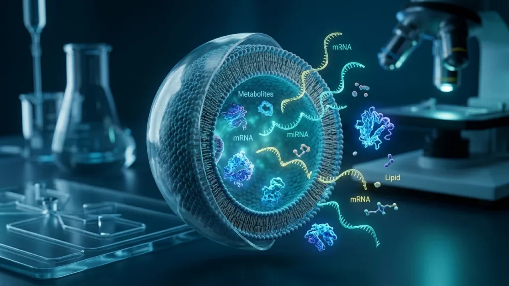
Exosomes are tiny packages. They are not simple bubbles. Their structure is precise and complex. This design lets them carry messages safely. So, what are exosomes made of? They have three key parts.
First, imagine a protective envelope. This is the outer membrane. It is a lipid bilayer. This means it is made of two layers of fat molecules. This membrane acts like a strong shipping box. It shields the precious contents inside. It also helps the exosome find the right cell to deliver its message. Specific molecules on its surface act like address labels.
Inside this protective envelope is the cargo. This is the most important part. The cargo is a mix of many different molecules. These molecules are the actual instructions or messages.
- They carry genetic blueprints like RNA.
- They include proteins that can change how a cell works.
- They may even contain bits of DNA or signaling fats.
The exact cargo depends on the parent cell. A healthy cell packs a normal message. A stressed or diseased cell might pack a harmful one. This cargo is why scientists are so interested.
The third critical component is the protein machinery. These proteins are embedded in the membrane or attached inside. They are the workers that make everything function. Some proteins helped form the exosome inside the cell. Others guide it to its destination. Special proteins fuse the exosome with a target cell. This fusion releases the cargo to do its job.
Think of it like a delivery drone. The outer hull is the membrane. The package inside is the cargo. The drone’s navigation system and landing gear are the protein machinery. All three parts must work together for a successful delivery.
This precise structure answers the question “what are exosomes made of?” It is not random. Every piece has a purpose. The lipid membrane provides safety. The inner cargo holds the information. The proteins enable delivery. This clever design allows exosomes to influence health and disease. Understanding these building blocks helps us see their true potential. Next, we will explore where these remarkable messengers come from and how cells make them.
How Exosomes Differ From Other Cell Vesicles
Cells release several kinds of tiny bubbles. Exosomes are just one type. They are often confused with their cousins, microvesicles and apoptotic bodies. Knowing the differences is crucial. It helps scientists know exactly what they are studying.
The first major difference is origin. Where do these vesicles come from? Exosomes form inside the cell. They start as inward buds in a compartment called the endosome. This creates a structure full of smaller vesicles. This is the multivesicular body. When it fuses with the cell’s outer membrane, it releases the exosomes. Think of it like a submarine launching mini-pods.
Microvesicles have a simpler origin. They bud directly from the cell’s outer membrane. It is like the cell pinching off a piece of itself. This process is faster and more direct.
Apoptotic bodies come from a dying cell. When a cell undergoes programmed death, it breaks apart. These bodies are the leftover fragments.
Size is another clear differentiator. You can think of it as small, medium, and large. – Exosomes are the smallest. They range from about 30 to 150 nanometers. That is thousands of times thinner than a human hair. – Microvesicles are larger. They range from 100 to 1000 nanometers. – Apoptotic bodies are the largest. They can be 1000 to 5000 nanometers across.
Their cargo also varies. This relates directly to what are exosomes made of. Exosomes carry a very specific set of molecules. Their cargo reflects their complex inner origin. It often includes specialized signaling proteins and genetic material like RNA.
Microvesicles carry cargo too. Their content can be more random. It often includes bits of the cell’s outer membrane and whatever was nearby when it budded.
Apoptotic bodies contain cellular debris. This can include whole organelles and chunks of DNA.
Finally, their roles in the body differ greatly. – Exosomes are messengers for communication. They send precise signals between healthy cells. – Microvesicles are often involved in immediate responses. For example, they help blood clot. – Apoptotic bodies are mostly for clean-up. They mark dead cell material for removal.
Why does this matter? In research, scientists must isolate pure exosomes. If a sample mixes exosomes with microvesicles, results get muddy. Understanding these differences ensures accurate science. It also highlights the unique design of exosomes. Their specific origin dictates their uniform size and rich cargo. This makes them ideal for sending complex messages throughout the body.
Now we know what they are and how they differ from other vesicles. The next logical question is about their journey. How do these precise messengers find their target cells to deliver their cargo?
Why Scientists Study These Tiny Packages
Scientists study exosomes because they are a new window into our health. These tiny packages act as biological reporters. They carry honest snapshots of the cells that made them. A diseased cell will pack a very different cargo than a healthy one. Researchers can analyze these differences. This gives them powerful clues about what is happening deep inside the body.
One major area is cancer research. Tumors use exosomes to their advantage. They send out exosomes that can prepare other parts of the body for cancer spread. These exosomes can suppress the immune system. They can also help create new blood vessels to feed a growing tumor. By studying these tumor exosomes, scientists learn how cancer works. They can also look for early signs of disease in a simple blood test. This is called a liquid biopsy.
Exosomes are also key players in brain health. In diseases like Alzheimer’s, harmful proteins can travel between brain cells inside exosomes. Studying this process helps explain how damage spreads. It also offers new targets for potential treatments. Researchers hope to one day block this bad communication.
The unique makeup of exosomes is central to this work. Understanding what are exosomes made of—their specific lipids, proteins, and RNA—is like finding a secret code. Each molecule has a story. For instance, certain surface proteins act like ZIP codes. They direct an exosome to a specific organ or cell type. This targeting is why they are so effective as messengers.
Their natural role makes them ideal for future therapies. Scientists are exploring how to engineer exosomes. The goal is to load them with helpful drugs or healing signals. Because our bodies already produce exosomes, they might cause fewer side effects than synthetic drug carriers. They could deliver medicine directly to sick cells.
Here are three concrete reasons researchers focus on these vesicles: – Early Detection: Exosomes in blood or urine can signal disease long before symptoms appear. – Understanding Disease: They reveal hidden communication pathways that cause illness. – Drug Delivery: Their natural design could be used for precise, targeted treatments.
The study of exosomes bridges basic biology and practical medicine. It turns cellular trash into valuable treasure. By decoding the messages in these tiny bubbles, scientists gain insights no scan can provide. This research moves us closer to a future of earlier diagnoses and smarter, more targeted therapies for many conditions. The next step is understanding how this knowledge is applied in real-world labs and clinics.
The Outer Shell: Understanding the Exosome Membrane
The Lipid Bilayer: Nature’s Protective Bubble
The exosome’s journey is a dangerous trip through the body’s fluids. Its protective shield is a lipid bilayer. This is a double layer of fat molecules. Think of it as nature’s own protective bubble wrap.
This bilayer is not a random sack. It has a precise structure. The molecules line up in two orderly rows. Their water-loving heads face outward. Their water-fearing tails hide inside. This creates a stable, flexible barrier.
This design is crucial for survival. The bilayer shields precious cargo from destruction. Enzymes and immune cells patrol bodily fluids. The membrane keeps them out. It ensures messages arrive intact at their destination.
The fats used are special. They are not simple storage fats. They are structural lipids like cholesterol and sphingomyelin. These molecules make the membrane sturdy yet fluid. They let the vesicle bend without breaking.
Membrane fluidity is key for fusion. An exosome must merge with a target cell to deliver its cargo. A rigid shell would fail. This flexible bubble can merge seamlessly. It hands off its package like a careful courier.
The bilayer also anchors critical tools. Proteins stick through it like gates and signposts. These are the “ZIP codes” mentioned earlier. They guide the exosome and help it communicate. Without the stable bilayer, these proteins would fall off.
So, what are exosomes made of at their core? Their foundation is this smart lipid wall. It is a delivery vehicle’s chassis. It provides safety, navigation, and the means for final delivery.
Consider these three key jobs of the lipid bilayer: – Protection: It creates a sealed compartment for fragile RNA and proteins. – Identity: Its specific lipid mix acts like a cellular fingerprint. – Delivery: Its physical properties enable the final handoff with a target cell.
Different cells use slightly different lipid recipes. A nerve cell’s exosome membrane differs from an immune cell’s. This affects where the vesicle can go and what it can do. The membrane defines the mission.
Understanding this shell solves a major puzzle. How can something so small travel so far? The answer lies in its durable, dynamic design. The lipid bilayer is more than a wall. It is an active part of the exosome’s message.
This protective bubble allows the precious cargo inside to remain secure. Next, we must look inside that bubble to see what it carries.
Membrane Proteins: The Address Labels of Exosomes
The exosome’s membrane is studded with special proteins. These are not random decorations. They are essential tools for navigation and communication. Think of them as the address labels and door handles of the tiny vesicle.
These membrane proteins have specific jobs. Some act as “ZIP codes.” They guide the exosome to the right neighborhood in the body. Others work like “keys.” They can unlock or bind to a target cell. This system ensures messages go only to the correct recipients.
The proteins are anchored firmly in the lipid bilayer. They stick through it. Their ends point both outward and inward. The outside parts interact with the world. The inside parts can signal to the cargo inside the exosome itself.
Let’s look at a few key types of these proteins: – Tetraspanins are very common. They form platforms. Other proteins gather around them. They help the exosome fuse with a target cell. – Integrins act like cellular glue. They decide which tissues an exosome will visit. An exosome with certain integrins may travel to bone, not liver. – Receptors are like antennas. They can pick up signals from the environment. This can tell the exosome when to deliver its cargo.
Cancer cells provide a clear example. They often put unique proteins on their exosomes. These proteins can help tumors spread. They might mark a spot in the body for a new tumor to grow. This shows how powerful these address labels can be.
So, what are exosomes made of that gives them direction? The membrane proteins are a major part of the answer. Without them, an exosome would be like a letter with no address. It would float aimlessly. Its important message would never be read.
Different cells equip their exosomes with different protein sets. A stem cell’s exosome has different labels than a skin cell’s exosome. This programming decides the vesicle’s fate and function.
The process is highly selective. A cell does not just dump proteins onto the exosome surface. It carefully chooses and places them during the exosome’s formation. This loading process determines the mission.
These proteins also help scientists identify exosomes. Researchers look for common surface proteins like CD9 or CD63. Finding these markers confirms they are studying a true exosome, not another vesicle.
In summary, the lipid bilayer provides the vehicle. The membrane proteins provide the map and the keys. They transform a simple bubble into a guided messenger. This targeting is why exosome research holds such promise for medicine.
The address system must be precise. A mistake could send a healing signal to the wrong cell type. The elegance lies in this biological specificity. The outside labels work in concert with the protected cargo inside.
Next, we will finally open the package. We will explore what these vesicles carry within their protective shell.
How the Membrane Stays Stable in Body Fluids
The exosome’s membrane is built for a rough journey. It must survive the bloodstream. It travels through other body fluids too. These liquids can be harsh. They contain salts and enzymes. They have forces that can tear fragile bubbles apart. Yet exosomes stay intact. Their stability comes from their special lipid wall.
Think of a soap bubble. It pops easily. An exosome is different. Its bilayer is not a simple soap film. It is a carefully built structure. The lipids are arranged in two layers. Their molecular shape provides natural strength. This double layer forms a stable barrier. It protects the precious cargo inside.
The specific lipids matter greatly. They are not random. Cells select certain types during exosome formation. These lipids have unique properties. – Some lipids are more rigid. They pack tightly together. – Others have special chemical groups. These groups repel water in a helpful way. – Cholesterol is often present. It acts like a molecular brace.
Cholesterol fits between other lipid molecules. This makes the membrane less fluid. It becomes more firm. This is crucial for stability in warm environments like the human body. Without it, the membrane might become too loose. It could leak or fall apart.
The lipid composition also prevents digestion. Body fluids contain enzymes called lipases. These enzymes break down fats. The exosome’s lipid mix is resistant to them. The arrangement of the lipids hides vulnerable bonds. This makes the vesicle durable over distance.
This durability answers part of what are exosomes made of. The answer includes these robust lipids. They form the primary shell. This shell gives the exosome its physical integrity. A weak shell would fail immediately. The message would never be delivered.
Temperature changes pose another challenge. The body maintains a steady temperature. But exosomes in research might be stored or shipped. Their membrane composition helps them handle mild shifts. The lipid bilayer can adapt without rupturing.
Surface charge adds another layer of protection. The outer face of the membrane often has a slight negative charge. This charge helps exosomes repel each other. It also helps them avoid sticking to certain structures prematurely. This keeps them circulating until they find their target.
The membrane’s strength is passive but vital. It does not require energy to maintain its form once released. This is a key engineering feat of nature. The cell builds a stable container in one efficient step.
In summary, the lipid bilayer is a resilient package. Its chemical design provides innate stability. This allows the address proteins on the surface to do their job. The cargo inside remains secure from release until delivery.
This robust design is why scientists can isolate exosomes from blood samples. The vesicles remain whole during processing. Their stability enables both their natural function and their study in labs. Next, we must look at what this stable shell is designed to protect: the core payload inside every vesicle.
Inside the Package: What Exosomes Carry
RNA Molecules: The Instruction Manuals Inside
The secure lipid bilayer protects a precious core. This core holds molecules called RNA. RNA is a type of genetic instruction. Cells use RNA to build proteins and control their own activity.
Exosomes carry different types of RNA. Each type has a specific job. The main types found inside are: – microRNA (miRNA). These are short strands. They do not build proteins. Instead, they silence genes. – Messenger RNA (mRNA). These are longer blueprints. They can be used to make new proteins. – Other regulatory RNAs. These have various control functions.
Think of these RNAs as software updates. A sending cell packages its current operational code. It then ships this code to another cell. The receiving cell downloads the new instructions. Its behavior can change as a result.
For example, a stem cell might send exosomes packed with specific miRNAs. These miRNAs can help a damaged cell switch on repair genes. The damaged cell might start healing faster. It did not need a direct stem cell touch. It just needed the instructions.
The process is precise and selective. Cells do not randomly dump RNA into exosomes. They carefully choose which molecules to pack. This selection determines the message’s effect.
Scientists study what are exosomes made of to understand this cargo. They find that an exosome from a heart cell differs from one from a brain cell. The RNA inside is tailored to its source.
Let’s look at microRNA more closely. These small molecules are powerful regulators. One miRNA can target hundreds of different messenger RNAs. It can shut down their signals.
This creates a wide network of influence. A single exosome delivering miRNAs can alter many pathways in a recipient cell. It can calm an overactive immune response. It can also slow down rapid cell growth.
Messenger RNA cargo is equally fascinating. If a recipient cell takes in an exosome with mRNA, it can use that blueprint. The cell’s machinery reads the new mRNA. It then produces a protein it might not normally make.
This protein could be a growth factor. It could be a specialized enzyme. This transfer expands the cell’s capabilities without changing its own DNA.
The packaging of RNA is also smart for stability. Naked RNA in the bloodstream would degrade quickly. Enzymes would chop it up instantly.
The exosome’s lipid bilayer shields the RNA during transit. The interior environment is also specially managed. This keeps the genetic instructions intact until delivery.
Research shows real-world impacts. Tumor cells send exosomes with unique RNAs. These RNAs can prepare distant organs for cancer spread. They can create a more welcoming environment for metastasis.
In contrast, healthy cells might send restorative signals. An exosome from muscle after exercise carries different RNAs than one from stressed muscle.
This cargo system explains why exosomes are such powerful messengers. They do not just signal with surface proteins. They deliver executable programs that rewrite cellular functions.
The recipient cell integrates these new instructions. It may change its metabolism. It might alter its growth rate. It could even start sending new signals of its own.
This transforms our view of cell communication. It is not just simple on-off signaling. It is the transfer of complex software between biological units.
Understanding this cargo is key for medicine. If we know the instructions, we might predict or change an outcome. We could intercept harmful messages. We could also design beneficial ones.
The next question is how these loaded packages know where to go. Their surface holds the addressing system for precise delivery.
DNA Fragments: Genetic Messages in Miniature
Exosomes carry more than just RNA instructions. They also transport pieces of DNA. This is not the cell’s full, perfect genome. Instead, exosomes contain fragments.
These DNA pieces come from two main sources inside the cell. The first source is the cell’s powerhouse, the mitochondria. Mitochondria have their own small circles of DNA. Exosomes can carry bits of this mitochondrial DNA.
The second source is the cell’s nucleus. This is where the main genome resides. Exosomes can contain fragments of this nuclear DNA too. The fragments are often double-stranded.
The DNA is protected within the exosome’s core. The lipid bilayer acts as a sturdy shipping container. This prevents the genetic material from degrading during its journey.
So, what are exosomes made of in terms of DNA cargo? The contents are a mixed snapshot. They reflect the cell’s state and even its history.
Researchers find this incredibly useful. Analyzing exosomal DNA provides a window into the cell of origin. It is a form of liquid biopsy. Scientists can detect mutations or damage without invasive surgery.
For example, tumor exosomes often carry telltale DNA. These fragments may contain the very mutations that drive the cancer. A recipient cell can take up this DNA.
This uptake can have profound effects. The foreign DNA fragments might integrate into the recipient cell’s genome. This is a rare but possible event. It could potentially alter the recipient cell’s behavior.
More commonly, the DNA acts as a signal. Specific sequences can trigger immune alerts. They might activate pattern-recognition receptors inside the receiving cell. This can spark an inflammatory response.
The process is highly selective. Cells do not randomly pack DNA into exosomes. Certain stressors influence what gets loaded. Damage or disease changes the profile.
Think of it like a cellular distress call or a broadcast. A stressed cell packages specific DNA fragments. It then sends them out for other cells to detect.
This adds a new layer to intercellular communication. It is not just protein signals or RNA programs. It is also the transfer of raw genetic data.
The implications for medicine are significant. Exosomal DNA could serve as an early warning system. Doctors might one day detect cancer from a simple blood draw. They would look for tumor-specific DNA in exosomes.
It also poses challenges for therapy. Harmful exosomes could spread damaging genetic material. Blocking this transfer might become a treatment goal.
Understanding this cargo completes the picture of what are exosomes made of. They are complex parcels with diverse contents. Each component, including these DNA fragments, serves a purpose in cellular dialogue.
The journey of these packages is equally precise. Their surface determines exactly which cells will receive their potent cargo.
Proteins That Do the Work Inside Cells
Proteins form the active workforce inside an exosome. They are the tools that directly change a recipient cell’s behavior. Think of DNA as an instruction manual. RNA might be a specific set of blueprints. Proteins are the actual builders and machines. They do the physical work.
These proteins are not random. The parent cell carefully selects them during packaging. This selection depends on the cell’s type and condition. A healthy cell sends routine maintenance signals. A stressed or diseased cell sends different commands.
Exosomal proteins fall into several key functional groups. Each group has a distinct job.
First are enzymes. These proteins speed up chemical reactions. An exosome from a liver cell might carry detoxifying enzymes. It could deliver them to a neighboring cell. This helps that cell break down harmful substances.
Second are signaling proteins. These molecules latch onto receptors on a target cell’s surface. This docking action triggers a chain reaction inside the cell. It is like turning a key to start an engine. Growth factors are a common example. They can tell a cell to grow, divide, or repair itself.
Third are structural proteins. They provide support and shape. Some help the exosome itself stay intact during its journey. Others can become part of the recipient cell’s internal skeleton after delivery.
Finally, there are regulatory proteins. These control gene activity. They can enter the nucleus of the target cell. Once there, they can switch other genes on or off. This changes the cell’s long-term program.
The source cell dictates the protein mix. An immune cell’s exosome will carry different proteins than a neuron’s exosome. The immune vesicle might contain inflammatory signals. The neural vesicle likely carries proteins for brain cell communication.
Cancer cells exploit this system brilliantly. Their exosomes are packed with specific proteins that help tumors grow. These proteins can break down local tissue to make space for the tumor. They can also shut down immune attacks. They even prepare distant organs for cancer spread. This process is called metastasis.
On the flip side, healthy cells use exosomal proteins for coordination. Stem cells release exosomes rich in healing proteins. These can encourage tissue regeneration and reduce scarring after injury.
Understanding this protein cargo answers a core part of what are exosomes made of. The lipids form the protective bubble. The nucleic acids carry information. The proteins provide immediate function. This combination makes exosomes powerful messengers.
Their journey does not end with delivery. The true impact happens when these proteins engage with the internal machinery of their new cellular home, directing its next actions with precision.
Lipids: More Than Just Membrane Material
The lipid membrane of an exosome is not just a simple bubble. It is a dynamic, organized structure. Its composition is highly specific. Cells carefully select the lipids they pack. This selection gives the exosome its identity and function.
Think of the membrane as a mosaic. It is made from many different lipid types. These include cholesterol, sphingomyelin, and ceramides. Each lipid has a unique job. Cholesterol provides stiffness and stability. Sphingomyelin helps form specialized areas called lipid rafts. These rafts are crucial. They act as assembly platforms for signaling molecules.
This specific lipid makeup answers part of what are exosomes made of. More importantly, it explains how exosomes work. The lipids themselves are bioactive signals. When an exosome fuses with a target cell, its lipids integrate into that cell’s own membrane. This transfer can change the recipient cell’s behavior immediately.
For example, ceramides can trigger programmed cell death. This is called apoptosis. Phosphatidylserine is another key lipid. It often sits on the inner layer of a cell’s membrane. When it flips to the outside, it sends an “eat me” signal. Immune cells recognize this signal. They then clear the marked cell or vesicle. Exosomes can carry phosphatidylserine on their surface. This helps control immune responses.
Lipids also serve as a concentrated energy source. The membrane is rich in fatty acids. These can be broken down for fuel by the receiving cell. This is especially important in stressful conditions. A starved cell might use exosomal lipids for immediate energy. This supports survival.
The lipid cargo has direct medical relevance. Cancer exosomes often have more cholesterol and sphingomyelin. This makes their membranes more rigid. This rigidity helps them survive longer in the bloodstream. It also protects their harmful cargo during transit to distant organs.
In contrast, exosomes from stem cells carry different lipids. These lipids can promote healing and reduce inflammation. They help create a better environment for tissue repair.
Here are three key roles of exosomal lipids: – They act as direct signals, changing recipient cell function. – They provide building blocks for new membranes in target cells. – They offer a ready supply of metabolic energy.
Researchers can analyze this lipid fingerprint. It tells them about the exosome’s origin and health status. A shift in lipid profiles can signal disease long before other symptoms appear. This makes lipids valuable biomarkers.
The journey of an exosome is powered by its dual cargo. Proteins execute precise commands. Lipids deliver foundational signals and fuel. Together, they ensure the message is not just delivered but also powered on arrival. This seamless integration highlights the sophistication of these biological packages, setting the stage for understanding their ultimate genetic instructions.
How Cells Make and Release Exosomes
The Birth of Exosomes Inside Cellular Factories
The creation of an exosome begins with a simple inward fold. The cell’s outer membrane curves inward. It forms a cup-like structure. This captures proteins and RNA from the surrounding cytoplasm. This pouch then pinches off inside the cell. It becomes an early endosome.
This early endosome is not yet an exosome. It is a sorting station. Its interior begins to change. The membrane of the endosome buds inward again. It creates tiny vesicles inside the larger one. This structure now looks like a bubble holding many smaller bubbles. Scientists call it a multivesicular body, or MVB.
The MVB is the true factory floor. Here, the cell makes critical choices. It decides what gets packaged into the tiny internal vesicles. Special protein complexes act as sorting machines. They recognize specific signal tags on cargo molecules. These machines guide lipids, RNA, and proteins into the forming vesicles.
This sorting answers the question what are exosomes made of. The cargo is not random. It is a precise snapshot of the cell’s state. A stressed cell will pack different cargo than a healthy one. A stem cell selects molecules for repair. A cancer cell may pack signals for growth.
The process relies on two main cellular pathways. The first is called the ESCRT pathway. This involves a team of proteins working together. They label the cargo. They help deform the membrane to bud inward. They then cut the vesicle free inside the MVB.
The second pathway is ESCRT-independent. Here, specific lipids in the membrane drive the process. Certain lipid types gather in specific areas. This causes the membrane to curve and form a vesicle on its own. Both pathways ensure tight packaging.
Once loaded, the vesicles float inside the MVB. The MVB itself must now decide its fate. It has two options. It can travel to and fuse with a structure called the lysosome. The lysosome is the cell’s recycling center. It will digest everything inside the MVB, including the small vesicles. This destroys the potential exosomes.
Alternatively, the MVB can travel to the cell’s outer membrane. It docks at the inner surface. The membranes of the MVB and the cell then fuse together. This opens the MVB to the outside world. The tiny internal vesicles are released into the extracellular space. At this moment, they earn their name: exosomes.
The cell controls this release decision carefully. Signals from outside the cell can guide it. More MVBs might be sent for release during injury or infection. The entire process, from first fold to final release, is fast. It can often complete within minutes.
This intricate manufacturing highlights a key point. Exosomes are not cellular waste. They are deliberate creations. Their birth is a regulated, multi-step assembly line. It ensures each tiny messenger is fully equipped for its journey outside the cell, ready to deliver its precise instructions to a waiting neighbor.
How Cells Choose What Goes Into Each Package
Cells do not pack exosomes at random. They carefully choose the cargo. This selection process answers a vital question: what are exosomes made of? The contents are not accidental. They are a deliberate snapshot of the cell’s state and intent.
Think of the forming exosome as a shipping container. The cell must decide what to put inside. It also decides what to attach to the outside walls. This loading happens during the inward budding stage described earlier.
The cargo falls into three main types. Each type has its own loading method.
First are membrane proteins. These get embedded directly into the exosome’s wall. Receptors and channels are examples. Their placement signals the exosome’s target. It is like putting a specific address label on the box.
Second is the internal cargo. This includes proteins, RNA, and even DNA fragments. These molecules float inside the exosome’s hollow center. They carry the actual instructions or tools for the recipient cell.
Third are lipids. The exosome membrane itself has a unique lipid mix. This composition aids in stability and fusion with target cells.
How does the cell sort this cargo? It uses molecular tags and sorting complexes. Many proteins destined for exosomes have a chemical tag. This tag is like a postal code. Cellular machinery recognizes this code. It guides the tagged protein into the forming vesicle.
For RNA molecules like microRNA, the process is also specific. Certain proteins bind to particular RNA sequences. These RNA-binding proteins often have tags that direct them into exosomes. So the RNA gets a piggyback ride into the package.
The cell’s condition directly controls the loading. A stressed cell will pack different cargo than a healthy one. For instance, a cancer cell might load exosomes with molecules that help tumors grow. An immune cell will pack signals to alert its neighbors during an infection.
This selectivity is powerful. It means exosomes from a heart cell will differ from exosomes from a brain cell. Even within one tissue, exosome content can change minute by minute. It reflects the cell’s immediate needs and messages.
Scientists can trace these sorting pathways. Disrupting them proves their importance. If you block a key sorting protein, the exosome’s cargo changes completely. Its message gets scrambled. This shows the system’s precision.
In summary, exosomes are made of a curated collection of biomolecules. Their composition is a direct result of active cellular selection. This turns each tiny vesicle into a targeted communication packet. The next logical question is: how do these precise packages know where to go and what to do upon delivery?
The Release Mechanism: Sending Messages Out
Once an exosome is fully loaded, it must exit the cell. This final step is called secretion. The cell does not simply let the vesicle go. It actively pushes the package out into the world.
The release mechanism often involves the cytoskeleton. This is a network of protein filaments inside the cell. Think of it as the cell’s scaffolding and transport system. Motor proteins walk along these filaments. They carry the mature exosome to the cell’s outer membrane.
Fusion with the plasma membrane is the critical event. The exosome’s own membrane touches the cell’s inner surface. The two lipid bilayers merge. This creates a temporary pore. The exosome’s contents are spilled directly into the extracellular space. The vesicle itself becomes part of the cell’s outer membrane for an instant before pinching off.
Several key proteins enable this fusion. They are called SNARE proteins. These proteins act like molecular zippers. One set is on the exosome membrane. The complementary set is on the cell’s inner membrane. They zip together. This pulls the membranes close and makes them fuse. Without SNAREs, exosomes would be trapped inside.
The rate of release is not constant. Cells control it tightly. Different signals tell a cell to increase or decrease exosome secretion.
- Cellular stress is a major trigger. Lack of oxygen, heat, or toxin exposure can cause a surge.
- Calcium levels inside the cell matter. A sudden rise in calcium often accelerates release.
- Signals from other cells can also command more secretion. An immune signal might tell a nearby cell to send out alert exosomes.
For example, a cancer cell secretes far more exosomes than a normal cell. This hyperactivity helps the tumor communicate. It can send signals to promote blood vessel growth or shut down immune attacks.
The extracellular space is not empty. It is filled with a gel-like substance called the matrix. Newly released exosomes navigate this dense meshwork. Some bind temporarily to matrix proteins. Others diffuse freely. Their journey to target cells begins immediately.
Understanding what exosomes are made of is only half the story. Their precise manufacture means nothing without this efficient delivery system. The release mechanism ensures the cellular message is sent on time. It completes the process from packaging to postage. The next mystery is how these messages find their exact recipient in the complex environment of the body.
The Journey of Exosomes Through the Body
How Exosomes Travel in Blood and Other Fluids
Once free from the tissue matrix, many exosomes enter the bloodstream. This is a major highway for long-distance travel. The blood is a busy and harsh environment. Exosomes must survive it to deliver their messages.
They do not travel alone. Most exosomes in blood are bound to carrier proteins. These proteins act like protective shields. They prevent exosomes from sticking to vessel walls prematurely. They also stop other blood components from breaking the exosomes down.
The journey is not random. Exosomes have address systems. Their surface acts like a shipping label. This label is made of the proteins and sugars stuck to their membrane. These molecules can dock with specific cells. This ensures a letter reaches the right mailbox.
Blood flow carries exosomes everywhere. Their small size is key here. At about 30 to 150 nanometers, they are far smaller than blood cells. This lets them travel through the tiniest capillaries. They can reach deep into tissues.
But how do they exit the bloodstream? They use a multi-step process.
First, an exosome slows down near its target tissue. The shipping label on its surface binds loosely to the vessel wall. This is like catching a railing from a moving boat.
Second, this bond triggers a stronger attachment. The exosome rolls to a stop.
Finally, the cell engulfs the vesicle. The exosome’s membrane fuses with the cell’s membrane. Its cargo is delivered inside. The message has been received.
Other body fluids also transport these vesicles. Exosomes are found in saliva, urine, and spinal fluid. In each fluid, they have different surface markers. These markers are suited for that specific environment. Salivary exosomes might target cells in the mouth or gut.
The stability of what exosomes are made of is crucial for this trip. Their tough lipid bilayer protects the cargo. It is resistant to enzymes in blood that would destroy a simpler packet. Without this robust construction, the message would degrade before arrival.
Travel distance varies widely. Some exosomes act locally on neighboring cells. Others cross the entire body. A vesicle released from a bone marrow cell can be found in distant liver tissue. This shows the scope of their reach.
Their concentration in blood is not trivial. Scientists estimate there are billions in a single milliliter of blood. This makes them powerful signalers. A disease like cancer changes this number dramatically. Tumors flood the system with their own exosomes.
This efficient transport solves a big biological problem. Cells need to talk to far-away organs. Hormones do this, but exosomes carry more complex instructions. They can deliver machinery, not just simple signals.
The bloodstream is a public network. Yet communication remains private because of precise addressing. The system’s elegance lies in this combination of broad distribution and specific delivery. Next, we must ask what happens after the message is opened inside the target cell.
Finding the Right Cell: The Delivery System
Exosomes do not bump into cells at random. Their delivery is a precise event. Think of it like a locked package. The exosome carries a set of keys on its surface. The target cell has the matching locks.
These “keys” are proteins and sugars. They stick out from the exosome’s lipid membrane. Scientists call them surface markers or ligands. Each type of cell in the body displays a unique set of “locks,” or receptors.
The match must be exact. An exosome from a nerve cell will have keys for other nerve cells or muscle cells. It will ignore a skin cell. This specificity ensures messages go only to the right address.
The process happens in clear steps. – First, the exosome circulates until it nears its target tissue. – Next, its surface keys bind tightly to the cell’s receptor locks. – This binding triggers the cell to pull the vesicle inside.
The entire structure of what exosomes are made of enables this. The lipid bilayer is a sturdy canvas. Proteins are embedded in this canvas like signposts. These signposts guide the exosome to its destination.
Some exosomes use a common key. A protein called CD63 is found on many vesicles. Others use very rare keys. A tumor exosome might display a marker only found on that cancer.
This system explains how an exosome from the brain can find a cell in the liver. The liver cell must express the correct receptor. If it does, the message is received. If not, the exosome moves on.
Research shows this is highly efficient. In lab tests, over 80% of certain exosomes bind to their intended target cells. The rest may be cleared by the immune system or break down.
The binding event is just the start. Once attached, the cell engulfs the vesicle. It pulls the exosome through its own membrane. The package is now inside the target cell’s territory.
The cargo remains protected until this moment. The tough bilayer shields it during transit. Now, inside the cell, the vesicle opens. Its molecular instructions are released.
This targeted delivery is vital for health. Immune cells send exosomes to coordinate an attack. Stem cells dispatch vesicles to repair damaged tissue. Each message finds its recipient.
Errors in addressing cause problems. In autoimmune disease, exosomes might target healthy tissue by mistake. In cancer, tumor vesicles can lock onto organs to spread disease.
The beauty lies in its simplicity. The system uses basic biological rules. It applies them with extraordinary precision. Billions of messages navigate our body daily. Nearly all find the correct door.
Understanding this code is a major scientific goal. If we learn the key-lock pairs, we could design smart therapies. We could send drug cargo directly to diseased cells.
The journey ends with successful delivery. The message has been transported and received. Next, we will see how the cell reads and acts upon these complex instructions.
How Exosomes Enter Cells to Deliver Messages
Once an exosome locks onto a cell’s surface, it must get inside. The cell does not simply open a door. It uses active processes to pull the vesicle in. Think of it as the cell reaching out to grab a delivered package.
The primary method is called endocytosis. The cell membrane folds inward. It wraps around the attached exosome. This forms a small pouch inside the cell. The pouch pinches off. Now the exosome is trapped in a bubble called an endosome. This is a common cellular import system.
There are several types of endocytosis. The specific type depends on the exosome’s surface markers.
- Clathrin-mediated endocytosis is a precise pathway. Proteins named clathrin form a coated pit on the membrane. This pit deepens and seals. It brings the exosome into the cell’s interior.
- Caveolin-mediated endocytosis uses different membrane proteins. These proteins create small flask-shaped invaginations. This route is common for signaling molecules.
- Macropinocytosis is like the cell taking a big gulp. Large sections of membrane ruffle and fold. They engulf fluid and anything attached, including exosomes.
Sometimes, exosomes use direct fusion. The lipid bilayer of the exosome merges with the cell’s own membrane. This is rare but efficient. It is like two soap bubbles becoming one. The exosome’s contents spill directly into the cell’s cytoplasm. No unpacking is needed.
The chosen path decides the cargo’s fate. Endocytosis often sends the exosome to an endosome. This compartment can become acidic. The change in pH can break open the vesicle. It releases the cargo safely inside the cell. Alternatively, the endosome might travel to other cell parts.
Direct fusion offers instant access. Messenger RNA or proteins enter the cellular workspace immediately. They can start influencing cell behavior without delay.
Cancer cells often exploit these entryways. Their exosomes may favor fusion or specific endocytosis types. This ensures rapid delivery of growth signals. It helps tumors manipulate their environment.
Immune cells use controlled endocytosis. They capture exosomes from pathogens or other cells. Then they analyze the cargo. This helps them identify threats and mount a defense.
The entry mechanism is not random. It is directed by what are exosomes made of. Their surface protein toolkit determines which cellular door they use. A different set of keys opens different locks.
Understanding these routes is crucial for medicine. Scientists can design therapeutic exosomes to choose a specific path. For a slow release, they might target endocytosis. For an immediate effect, they could engineer vesicles for fusion.
The journey inside is complete once the cargo is freed. The protective lipid bilayer has done its job. The molecular messages are now in the right hands. Next, the cell must decode and act on these complex instructions, turning signals into action.
Exosomes in Health: The Normal Communication System
How Healthy Cells Use Exosomes to Talk
Healthy cells are constantly chatting. They send thousands of exosomes every day. This traffic is not a sign of disease. It is essential for a body to function as one coordinated system.
Think of a tissue as a neighborhood. Cells are the houses. Exosomes are the mail trucks. They deliver precise instructions and supplies. This keeps the entire community stable and responsive.
The immune system relies heavily on this mail service. When a cell detects a virus, it does not just scream for help. It packages viral fragments into exosomes. These vesicles then travel to immune cells nearby. They deliver the evidence. This is like handing a wanted poster directly to the police. It helps activate a targeted defense much faster.
Exosomes also carry orders to calm down. After an infection is cleared, some cells send anti-inflammatory signals. Their exosomes tell immune cells to stop attacking. This prevents damage to healthy tissue. It is a crucial “cease-fire” command.
Beyond defense, exosomes are master coordinators for repair. After an injury, stem cells and other repair cells release special vesicles. Their cargo includes growth factors and instructions for rebuilding. These exosomes travel to the damage site. They tell local cells to multiply. They guide them to form new blood vessels. They help lay down fresh collagen for healing.
This repair toolkit is part of what are exosomes made of. Their lipid membrane protects the delicate growth signals during transit. Their surface proteins help them find the exact cells that need the instructions.
Another vital job is waste removal. Cells constantly renew their machinery. Old or misfolded proteins must be cleared out. The cell can package this waste into exosomes. It then ships the vesicles out for disposal. This keeps the cell’s interior clean and functional. Neighboring cells or systemic filters can then break down this material.
In the brain, neurons use exosomes for maintenance and signaling. They send vesicles along long neural pathways. These can deliver nutrients to supporting cells. They may also help prune unnecessary connections. This keeps the neural network efficient.
Even everyday metabolism is guided by exosomes. Fat cells, for instance, release vesicles that influence how the body handles sugar and insulin. Liver cells send out exosomes that affect cholesterol processing in other tissues. This constant exchange helps maintain metabolic balance.
The key is that this communication is precise and local. The messages are packaged for specific recipients. The system is elegant and efficient. It avoids broadcasting signals to the whole body, which would cause confusion.
When this system works well, tissues stay healthy. Organs coordinate their activities seamlessly. The body maintains a state of balance, or homeostasis. This quiet, constant dialogue is the foundation of our normal physiology.
Disrupting this talk can lead to disease. But understanding it gives science a blueprint. Researchers can learn how to copy these natural messages for therapy. They can design treatments that speak the body’s own language to promote healing from within.
Exosomes in Immune System Coordination
The immune system is a vast network of cells spread throughout your body. These cells must communicate quickly to find and stop threats. Exosomes are their instant messaging system. They carry urgent instructions between distant immune cells.
Imagine a scout cell patrolling your skin. It finds a potential invader, like a virus. The scout does not fight alone. It immediately releases exosomes loaded with specific signals. These vesicles travel through bodily fluids. They seek out helper T-cells and other immune soldiers.
What are exosomes made of in this case? Their cargo is the message. It includes pieces of the invader, called antigens. It also contains activation codes, like proteins called cytokines. This precise packaging tells receiving cells exactly what to look for and how to respond.
The arriving exosomes dock onto a helper T-cell. The T-cell reads the cargo. It now knows the enemy’s identity. The T-cell becomes activated. It starts to multiply, creating an army of clones specifically tuned to that virus.
These cloned T-cells then use their own exosomes. They send out orders to other parts of the immune system. Their vesicles can do several key things: – Direct B-cells to make antibodies that stick to the virus. – Rally natural killer cells to destroy infected body cells. – Signal macrophages to come and clean up the debris.
This coordination is fast and local. It prevents a full-body alarm that could cause harmful inflammation. Exosomes allow for a targeted strike instead of a blanket bomb.
The communication does not stop when the threat is gone. Regulatory T-cells send calming exosomes once the battle is won. These vesicles carry signals that tell the aggressive cells to stand down. They help turn off the immune response. This prevents the immune system from accidentally attacking your own healthy tissues.
Without this exosome dialogue, the immune system would be chaotic. Cells would not know when or where to attack. They might not stop attacking after the fight. This precise vesicle traffic keeps defense efficient and safe.
Researchers see great promise here. By studying these natural immune messages, they could design new therapies. Future treatments might use engineered exosomes to train the immune system against cancer. They could also use them to calm an overactive immune system in autoimmune diseases.
This shows how health relies on flawless cellular talk. The immune system’s power depends not just on strong cells, but on their perfect coordination. Exosomes provide the critical link for that teamwork, ensuring defense is both powerful and precise.
Tissue Repair and Maintenance Messages
Healthy tissues are not static. They constantly renew and repair themselves. This ongoing upkeep relies on clear communication. Exosomes provide the messaging system for this daily maintenance. They carry the precise instructions needed for tissue repair.
So, what are exosomes made of? Their structure is key to their role. Each exosome is a tiny bubble wrapped in a protective lipid membrane. This membrane shields precious cargo during transit. Inside, they carry proteins and genetic instructions. These instructions tell recipient cells what to do.
Consider a small cut on your skin. The repair process starts immediately. Cells near the injury release special exosomes. These vesicles travel to nearby stem cells. They deliver growth factors and RNA messages. The stem cells get the signal to multiply and move to the wound site. They then differentiate into new skin cells to close the gap. This exosome-driven process is fast and local. It ensures repair happens exactly where it is needed.
Beyond emergency repair, exosomes manage routine renewal. Your bone tissue is constantly being reshaped. Old bone is broken down by cells called osteoclasts. New bone is built by cells called osteoblasts. These two cell types must work in perfect balance. Exosomes coordinate their activity. Osteoblasts send exosomes to control osteoclast formation. This keeps bone removal in check. Without these messages, bone density could be lost.
The same principle applies to organ health. In the liver, exosomes help detoxify the body. Liver cells send vesicles containing enzymes to neighboring cells. This shares the metabolic workload. It prevents any single cell from being overwhelmed. In the brain, neurons release exosomes to support surrounding glial cells. This maintains the health of the neural network.
The cargo inside these maintenance exosomes is carefully selected. It often includes: – Growth factors that stimulate cell division. – Enzymes that help break down waste products. – MicroRNAs that regulate gene expression in the target cell. – Structural proteins that help rebuild the extracellular matrix.
This cargo is not random. The parent cell packages it based on the tissue’s current needs. A stressed cell will send different signals than a resting cell. The recipient cell accepts the exosome and unloads its instructions. It then changes its behavior to support tissue health.
This system is remarkably efficient. It allows for a decentralized response. There is no single command center directing every repair job. Instead, each cell can send and receive vital updates. This creates a resilient and adaptive network. Problems can be addressed locally before they become systemic.
The constant flow of these vesicles is a background process of health. It is less dramatic than an immune battle but equally vital. While immune exosomes respond to threats, maintenance exosomes prevent them. They keep tissues strong and functional every day.
This seamless communication is the foundation of resilience. When it works well, we experience it as uninterrupted health. The next section will explore what happens when this delicate system is disrupted.
When Communication Goes Wrong: Exosomes in Disease
How Cancer Cells Hijack Exosome Systems
Cancer cells are not just growing out of control. They are master manipulators of biological communication. A single tumor can release up to ten times more exosomes than normal tissue. This flood of vesicles becomes a powerful tool for invasion and survival.
So, what are exosomes made of when they come from a cancer cell? Their cargo is carefully packed to help the tumor. It is a malicious toolkit designed to reshape the environment. This cargo allows the cancer to spread long before any cell physically moves.
First, tumor exosomes prepare new ground. They travel to distant organs. Their signals make those areas more welcoming for cancer cells. They do this by breaking down local structures and promoting blood vessel growth. This process is called creating a pre-metastatic niche. It is like sending advance scouts to set up camp.
Second, they suppress the immune system. The exosomes carry molecules that confuse or deactivate immune cells. They can tell T-cells to stand down. They can reprogram macrophages into helpers that protect the tumor. This lets the cancer grow hidden from the body’s defenses.
Third, they spread treatment resistance. Exosomes can absorb chemotherapy drugs and carry them away from the tumor. They also send genetic instructions to other cancer cells. These instructions tell them how to survive the drug attack. This makes the entire tumor harder to kill.
The contents of a typical tumor exosome include: – Oncogenic proteins that force normal cells to grow. – MicroRNAs that shut down tumor-suppressor genes in healthy cells. – Enzymes that chew through tissue barriers, clearing a path for invasion. – Ligands that directly turn off immune cell activity.
This hijacking has devastating effects. The constant stream of bad information corrupts the local tissue network. Healthy cells around the tumor start helping it instead of fighting it. The corrupted exosomes also travel through the bloodstream. They can affect organs far from the original cancer site.
The result is a self-sustaining cycle. The tumor uses exosomes to grow and protect itself. Its growth leads to more tumor cells. More tumor cells send even more malicious exosomes. This cycle accelerates disease progression.
Understanding this hijack is crucial. It shows that cancer is a systemic communicator, not just a localized lump. The disease spreads its influence through biological messages in vesicles. This knowledge shifts how we view metastasis and resistance. It is not just about cells moving. It is about information moving first.
This corrupted communication system presents both a challenge and an opportunity for medicine. The next logical step is to explore how science is learning to intercept these messages or even turn this system against the disease itself.
Exosomes in Neurological Disorders
Exosomes play a key role in neurological diseases like Alzheimer’s. In a healthy brain, cells use exosomes to share helpful signals and remove waste. In Alzheimer’s, this system breaks down. The problem starts with two faulty proteins. These proteins are called amyloid-beta and tau.
Amyloid-beta proteins can clump together outside neurons. Tau proteins tangle up inside them. These clumps and tangles are toxic. They disrupt communication between brain cells. Eventually, they lead to cell death. This causes memory loss and confusion.
So how do exosomes fit in? Brain cells package these harmful proteins into exosomes. Think of it as a cell mistakenly mailing out a dangerous item. The exosome’s lipid bilayer membrane protects the cargo. This allows it to travel safely to a neighboring cell.
The receiving cell accepts the exosome. It then unpacks the toxic proteins inside itself. This spreads the damage. The process turns a local problem into a widespread one. Exosomes act as delivery vehicles for disease.
Scientists have found evidence in patient samples. The exosomes from Alzheimer’s patients contain high levels of amyloid-beta and tau. This is part of what exosomes are made of in this disease state. Their cargo is dangerous.
This spread follows a specific pattern in the brain. It often matches the path of disease progression. The exosomal spread helps explain why Alzheimer’s starts in one area and moves to others.
But exosomes are not all bad in this story. They have a dual role. Cells also use exosomes to try and clear away these toxic proteins. It is a disposal mechanism. Sometimes the system gets overwhelmed. The garbage removal fails, and the harmful cargo gets sent out instead.
Researchers are studying this closely. They want to find the exact moment the switch happens from cleanup to spread. Understanding this could lead to new treatments.
Potential therapeutic ideas are emerging. One idea is to stop exosomes from carrying toxic proteins. Another is to guide exosomes to carry helpful drugs instead. Scientists could design exosomes to deliver enzymes that break down the clumps.
The story of exosomes in Alzheimer’s is complex. They are part of the problem and a potential solution. This mirrors their role in cancer. In both cases, a natural messaging system becomes corrupted.
The focus now shifts to the body’s defense network. How does the immune system normally interact with these tiny vesicles? This interaction is critical for health and disease.
Inflammatory Diseases and Faulty Messages
Inflammatory diseases often involve a case of mistaken identity by the immune system. The body’s defenses attack its own healthy tissues. Exosomes play a central role in this faulty communication. They can carry and spread the very signals that turn peace into war.
Consider rheumatoid arthritis. In this disease, the lining of the joints becomes inflamed and painful. Cells in this lining, called synovial fibroblasts, release large numbers of exosomes. These vesicles are not empty packages. Their cargo is specific and damaging.
So, what are exosomes made of in this context? Their load includes potent inflammatory molecules. – They carry cytokines, which are chemical alarm signals. – They transport enzymes that break down tissue. – They present self-antigens that confuse immune cells.
This cargo transforms the exosome into a pro-inflammatory bullet. When released, it travels to nearby immune cells. An immune cell like a macrophage receives the exosome. It absorbs the vesicle and reads its contents. The inflammatory instructions inside activate the cell.
The activated macrophage then releases its own barrage of signals. This creates a vicious cycle. More inflammation triggers more exosome release. More exosomes activate more immune cells. The joint becomes a battlefield of constant, misguided attack.
The process is not limited to arthritis. Similar exosomal messaging occurs in lupus and inflammatory bowel disease. In lupus, exosomes from damaged cells carry nuclear proteins. These proteins look like threats to the immune system. This prompts a widespread autoimmune reaction.
Researchers have measured these effects. Exosomes from patients with active disease can directly cause inflammation in lab models. Healthy cells exposed to these exosomes start acting like diseased cells. This proves the vesicles are not just bystanders. They are active drivers of pathology.
The lipid membrane of the exosome is key here. It protects the inflammatory cargo during transit. This ensures the signals arrive intact and potent. The membrane also determines which cells receive the message. Specific surface molecules act like address labels, targeting immune cells precisely.
Blocking this communication is a major research goal. Scientists are testing ideas to intercept faulty exosomes. – One approach uses molecules to bind and neutralize them in circulation. – Another tries to stop cells from packaging harmful cargo in the first place. – A third strategy aims to edit the exosome’s address label, misdirecting it.
Understanding this mechanism reframes chronic inflammation. It is partly a disease of cell-to-cell messaging. Tiny vesicles amplify and perpetuate the wrong signals. Stopping the disease may require jamming this biological broadcast system.
This reveals a common thread across different illnesses. From Alzheimer’s to arthritis, corrupted exosomal communication worsens disease progression. The next logical question involves control. If these vesicles can spread harm, can we also engineer them to spread healing? This potential turns them into tools for therapy.
Medical Applications: Using Exosomes for Health
Exosomes as Disease Detection Tools
Doctors can now find disease clues in a simple blood draw. These clues are exosomes. Sick cells, like cancer cells, release far more exosomes than healthy ones. They also pack them with different cargo. This cargo reflects the cell’s exact state. Think of it as a molecular snapshot mailed into your bloodstream.
This makes exosomes powerful detection tools. They offer a “liquid biopsy.” This is less invasive than cutting tissue. A standard biopsy looks at cells stuck in one place. Exosomes provide information from cells throughout the body. They are especially useful for finding hard-to-reach tumors.
So, what are exosomes made of that allows this? Their structure is key. The protective lipid bilayer shields the internal cargo from degradation. Inside, scientists find specific molecules that signal disease. – MicroRNAs can show if a cell is becoming cancerous. – Faulty proteins may hint at early brain disease. – Unique surface markers act like a return address, pointing to the cell of origin.
For example, pancreatic cancer is often found too late. Researchers discovered that exosomes from these tumors carry a specific protein signature. This signature appears in blood long before symptoms do. Finding it early could dramatically improve survival.
The process for using them is straightforward. First, a blood sample is taken from a patient. Next, exosomes are separated from other blood components. Then, their cargo is analyzed for known disease markers. This search can be done for a single disease or many at once.
The advantages are clear. Testing is minimally invasive and can be repeated often. It provides real-time updates on a disease’s status. Monitoring exosome levels can also show if a treatment is working. A drop in harmful exosomes might mean therapy is succeeding.
This technology is moving fast. Several diagnostic tests using exosomes are in clinical trials. They target cancers, neurodegenerative diseases, and liver conditions. The goal is to catch illness at its most treatable stage.
This shifts exosomes from threat to asset. Their role as precise messengers becomes a medical advantage. We can intercept and read their mail for early warnings. This turns the body’s communication system into a built-in alarm. The next step is using that knowledge not just to detect, but to treat.
Therapeutic Exosomes: Nature’s Drug Delivery System
Exosomes are not just messengers. They can also become tiny treatment trucks. Scientists are learning how to load them with medicine. The goal is to send these drugs straight to sick cells. This method could be better than standard treatments. It aims to be more precise and cause fewer side effects.
So, what are exosomes made of? Their structure is key to this job. They have a protective lipid bubble. This bubble is like a cell’s own membrane. The body recognizes this as natural. It does not attack it quickly. This lets exosomes travel safely in blood. Their surface holds address proteins. These proteins guide the exosome to certain cell types.
Think of an exosome as a smart package. The outer envelope is the shipping box. The address label is the protein on the surface. The medicine inside is the cargo. The system uses the body’s own mail routes.
Loading the cargo is a major research area. Scientists use several methods. – They can incubate exosomes with drug molecules. The drugs slowly seep inside. – They use electrical pulses to temporarily open the exosome’s membrane. Medicine is pushed in during this opening. – They can even engineer parent cells. They make the cells produce exosomes that already contain the treatment.
A key example is cancer therapy. Chemotherapy drugs often harm healthy cells. This causes severe side effects. Researchers pack these drugs into exosomes. They then attach special markers to the exosome’s surface. These markers match proteins found only on the tumor. The exosome delivers its toxic cargo directly to the cancer. It largely avoids other tissues.
This approach is also promising for brain diseases. The brain has a protective shield called the blood-brain barrier. Most drugs cannot cross it. Exosomes from certain cells, like stem cells, can naturally cross this barrier. They could carry drugs for Alzheimer’s or Parkinson’s disease right to the brain cells that need help.
The advantages of this system are significant. – It uses a natural delivery vehicle. The body tolerates it well. – It protects fragile treatments. The lipid bilayer shields mRNA or proteins from degradation. – It allows for targeted action. Treatments go where they are needed most.
Current research is very active. Early studies show success in lab animals. For instance, exosomes loaded with anti-cancer drugs shrank tumors in mice. Other experiments delivered gene therapy tools to repair damaged cells.
Challenges remain. Scientists must control how much drug each exosome carries. They need to ensure exosomes go exactly to the right target every time. Production of medical-grade exosomes must be scaled up safely.
The vision is powerful. One day, a patient’s own cells could be used to create custom exosomes. These would be filled with personalized medicine. Then they would be injected back into the body to heal from within. This turns our cellular communication network into a therapeutic highway, moving us from detection to direct repair.
Regenerative Medicine and Tissue Healing
Exosomes carry natural instructions for repair. They are not just empty bags for drugs. Their own cargo can tell damaged cells to heal themselves.
This is the core idea behind regenerative medicine. The goal is to fix damaged organs. The body often cannot do this completely after a major injury. Exosomes could provide the needed signals.
So, what are exosomes made of that gives them this power? Their contents are key. They contain growth factors and instructions called microRNAs. These molecules can switch on repair programs in target cells.
Think of a damaged heart after a heart attack. Scar tissue forms. This weakens the muscle. Researchers have used exosomes from stem cells in experiments. These exosomes travel to the injured heart tissue.
They deliver their cargo to the surviving heart cells. The microRNAs inside tell these cells to grow new blood vessels. This process is called angiogenesis. Better blood flow brings more oxygen and nutrients. This helps the heart muscle recover and reduces scarring.
The same principle applies to bones and tendons. A severe fracture might not heal well. Exosomes from mesenchymal stem cells are being studied. They can encourage new bone formation. They guide stem cells at the injury site to become bone-building cells called osteoblasts.
For chronic wounds, like diabetic ulcers, healing is often slow. Exosomes can promote skin repair. They signal skin cells to multiply and move. They also reduce harmful inflammation at the wound site.
The advantages of using exosomes for regeneration are clear. – They are natural signaling packages. The body already uses them for communication. – They seem safer than whole cell therapies. Exosomes cannot multiply or form tumors. – They have a complex cargo. They carry hundreds of healing molecules at once. This is hard to replicate with a single drug.
One promising area is nerve regeneration. Damage to nerves in the spine or limbs is often permanent. Exosomes from support cells in the nervous system show potential. In lab studies, they help nerves regrow over gaps.
They do this by creating a supportive environment. The exosomes supply proteins that act as a guide rail for growing nerve fibers. They also calm the immune response that can block repair.
The future could involve off-the-shelf exosome products for specific injuries. A standardized exosome preparation for muscle tears is one possibility. Another is a gel infused with exosomes for applying directly to burns.
The vision extends to organ repair without major surgery. Imagine an injection of exosomes helping a damaged liver regenerate its own tissue. This approach uses the body’s own language to trigger healing.
It moves beyond simply slowing disease. It aims for true restoration of function. This represents the next frontier in using our biology to fix itself. The journey from understanding these tiny vesicles to applying them as healers is now underway.
The Future of Exosome Research and Medicine
Current Challenges in Exosome Science
Scientists are excited about exosome medicine. But major hurdles remain. These tiny messengers are complex. Turning them into reliable treatments is not simple.
One big question is about their origin. What are exosomes made of changes with their source. Exosomes from a stem cell differ from those of a cancer cell. Their cargo depends on the parent cell’s health and environment. This is a problem for making consistent medicine. A batch made on Monday must match a batch made on Friday.
Standardizing production is a huge technical challenge. Isolating pure exosomes is difficult. They are incredibly small. Current methods often collect a mix of vesicles and other particles. Contaminants can skew results. Scientists need better tools to sort exosomes from similar-looking bubbles.
Scaling up presents another obstacle. Growing enough cells to harvest exosomes for thousands of patients is costly. The purification process must be efficient at large volumes. It must also keep the exosomes intact and functional. A damaged lipid bilayer ruins the package.
Delivery is a critical puzzle. How do you get exosomes to the right place? Injecting them into the bloodstream seems direct. But many get filtered out by the liver or destroyed. Scientists are testing protective coatings. They are also exploring local delivery. This means applying exosomes directly to a wound or a diseased joint.
Tracking exosomes inside the body is another issue. We need to know where they go. Do they reach the target tissue? How long do they stay? Advanced imaging techniques are being developed for this. They use safe tags that glow under special scanners.
Regulation and safety monitoring need new frameworks. Exosomes are not traditional drugs. Agencies are creating guidelines for their approval. Long-term effects must be studied carefully. Even though exosomes are natural, delivering a large concentrated dose is new.
The cost of future therapies is uncertain. Complex manufacturing could make treatments expensive initially. The goal is to develop processes that eventually bring costs down.
Finally, we must fully decode their language. An exosome carries hundreds of signals. Scientists are still mapping which molecules do what. Understanding this communication network is key. It will allow us to design even smarter exosome-based treatments.
Solving these challenges requires collaboration across fields. Biologists, engineers, and clinicians must work together. Each solved problem brings us closer to safe and effective exosome medicine. The path forward is clear but requires careful, step-by-step progress.
Promising Research Directions for Tomorrow
Scientists are now designing exosomes in the lab. They can load these vesicles with specific drugs or genetic instructions. This turns natural messengers into targeted delivery trucks. The goal is to hit diseased cells with precision. This approach could reduce side effects common in current treatments.
One major direction is cancer research. Tumors release many exosomes to spread. Researchers are flipping this script. They are creating exosomes that carry anti-cancer signals. These engineered vesicles might tell cancer cells to stop growing. They could also deliver chemotherapy directly inside tumors. Early tests in models show this can shrink cancers with lower drug doses.
Another exciting area is brain diseases. The blood-brain barrier protects the brain. It also blocks most medicines. Exosomes can cross this barrier naturally. Scientists are using this ability. They are packing exosomes with therapies for Alzheimer’s or Parkinson’s. The vesicles ferry the treatment to the brain’s neurons. This was nearly impossible before.
Regenerative medicine is also advancing. The question of what are exosomes made of is key here. By understanding their cargo, scientists can boost healing. For instance, they can isolate exosomes rich in growth factors from stem cells. These exosomes could then repair heart tissue after a heart attack. They might also regrow damaged tendons without surgery.
Diagnostics is a parallel field. Doctors could use exosomes as early warning signals. A simple blood draw could provide a “liquid biopsy.” Exosomes from a tumor carry its unique markers. Finding these vesicles might catch cancer years earlier than a scan can. This research focuses on sensitive detection tools.
Personalized medicine is the ultimate horizon. Your own cells could produce therapeutic exosomes. A doctor might take a sample of your skin cells. These cells would be reprogrammed in a lab to make healing exosomes. Then those personalized vesicles would be given back to you. This could minimize immune reactions.
Key research tools driving this future include: – Advanced imaging to watch exosomes work in real time. – Microfluidic chips to sort and analyze exosomes faster. – AI models to decode the complex signals in exosome cargo. – New methods to scale up production for clinical use.
The path is moving from observation to engineering. Researchers are not just studying exosomes anymore. They are actively building and directing them. This shift marks a new phase in medical science. It promises more intelligent and less invasive therapies for many conditions. The next decade will likely see these research directions move into clinical trials, bringing theory closer to real-world help.
How Understanding Exosome Composition Changes Medicine
Knowing exactly what exosomes are made of changes everything in medicine. It turns these tiny vesicles from mysterious bubbles into precise medical tools. Their composition is the key. This knowledge allows doctors to intercept bad messages and send good ones.
Think of a harmful exosome as a disguised enemy truck. It carries specific cargo to help disease spread. Scientists can now read the shipping manifest on that truck. They can see the unique proteins on its surface. This tells them exactly where the truck came from. A tumor exosome has different surface markers than one from a healthy cell. This is the basis for new blood tests. Doctors can find these dangerous vesicles long before a tumor forms a visible lump.
Understanding cargo leads to smarter treatments. Researchers can now engineer exosomes. They load them with specific therapeutic molecules. This is like packing a rescue vehicle with exactly the right supplies. The lipid membrane of the exosome acts as a natural delivery capsule. It protects the medicine until it reaches the target cell. This method is more efficient than injecting drugs alone. The drugs are less likely to get lost or break down.
Medicine becomes highly targeted. Knowing the surface proteins lets scientists steer exosomes. They can add special tags to the vesicle’s membrane. These tags act like GPS coordinates. They guide the exosome to liver cells, brain cells, or heart cells. This precision reduces side effects. Healthy cells are left alone while sick cells get the treatment.
The impact spans across different diseases: – In neurodegeneration, engineered exosomes might carry growth factors to repair brain cells. – For autoimmune disorders, they could deliver signals to calm an overactive immune system. – In wound healing, exosomes from stem cells can instruct skin cells to regenerate faster.
This shift is fundamental. Medicine is moving from treating symptoms to reprogramming cellular communication. The focus is on information and delivery. Researchers decode the biological messages in exosome cargo. Then they rewrite them for healing. This approach is less invasive than major surgery. It is more targeted than broad-acting drugs.
The future of this field relies on continued mapping. Scientists are still cataloging all the parts inside different exosomes. Each discovery adds a new tool to the medical kit. The goal is a comprehensive library. A doctor could match a patient’s exosome profile to a specific condition. Then they could choose a pre-designed therapeutic exosome to correct it.
This transforms healthcare from reactive to proactive and precise. It turns our body’s own messaging system into a powerful ally for healing. The next step is refining these tools for safe, widespread use in clinics everywhere.
Your Takeaway: Why Exosome Structure Matters for Everyone
Key Points About What Exosomes Are Made Of
Think of an exosome as a tiny biological package. Its structure is what makes it so special. This structure allows it to carry messages and protect its cargo. It is not a simple bubble. It is a sophisticated, engineered vessel.
So, what are exosomes made of? They have three main parts. The first part is the outer membrane. This membrane is a double layer of fats, or lipids. This lipid bilayer acts like a strong envelope. It shields the contents inside from the harsh environment outside the cell.
The membrane is not smooth. It is studded with many different proteins. These proteins are crucial. Some act like name tags. They tell other cells where the exosome came from. Others work like docking ports. They help the exosome latch onto a specific target cell.
The second major part is the cargo inside. This cargo floats in the liquid center. It is a rich mixture of functional molecules. The cargo includes microRNAs. These are small pieces of genetic code that can silence genes in a recipient cell. It also includes other RNA types, proteins, and sometimes even DNA fragments.
This cargo is not random junk. It is carefully selected by the parent cell. A healthy cell packs a different cargo than a stressed cell. A cancer cell packs a very different cargo than a normal cell. The cargo reflects the cell’s state and its intended message.
The third key element is the machinery. This includes proteins used for making, loading, and releasing the vesicle. Even after release, these proteins can help the exosome function. They might help fuse with a target cell or unload the cargo.
Understanding this composition solves a mystery. It explains how exosomes work. The membrane proteins guide delivery. The lipid bilayer protects the fragile molecules inside during transit. The active cargo changes the behavior of the cell that receives it.
Each component has a direct impact on potential medical uses. Scientists can engineer each part. – They can change membrane proteins to improve targeting. – They can load the vesicle with therapeutic drugs or healing RNA. – They can use lipids from certain cells to make vesicles more stable.
This is why knowing what exosomes are made of matters for everyone. It is the blueprint for their natural role in health and disease. It is also the blueprint for future medicines. If we know the parts, we can repair or improve them. We can build custom versions for specific tasks.
For example, exosomes from stem cells are packed with growth factors. These factors help tissues regenerate. Exosomes from a tumor, however, are packed with signals that help cancer spread. The difference is entirely in their composition.
The takeaway is clear. The power of an exosome lies in its precise construction. Its makeup defines its journey through the body. Its makeup defines its message. By learning this biological language of lipids and proteins, we learn to speak it for healing. This fundamental knowledge turns these natural particles into tools we can truly use.
How This Knowledge Affects Future Health Care
Understanding what exosomes are made of opens direct paths to new medical tools. This knowledge moves us from observation to action. We can now design these natural carriers for specific health problems.
One clear goal is targeted drug delivery. Many powerful drugs harm healthy cells. This causes severe side effects. Exosomes offer a smarter solution. Scientists can load a drug inside the vesicle. They can then engineer the exosome’s surface proteins. These proteins act like a postal code. They guide the vesicle only to sick cells, such as cancer cells. The lipid bilayer safely carries the drug through the bloodstream. It then delivers its cargo directly to the target. This method could make treatments more effective. It could also make them much safer for patients.
Diagnostics will also change dramatically. Exosomes from different cells carry unique molecular signatures. A simple blood test could analyze these vesicles. Doctors could detect diseases like cancer or Alzheimer’s very early. They could do this long before symptoms appear. Early detection often leads to better outcomes. This approach is often called a “liquid biopsy.” It is far less invasive than tissue sampling.
The field of regenerative medicine will benefit too. Our bodies use exosomes for natural repair. Stem cell exosomes, for instance, carry instructions for healing. Scientists are learning to harvest or mimic these vesicles. They could be used to treat injured tissues. – Damaged heart muscle after a heart attack could be prompted to repair itself. – Arthritic joints could receive signals to reduce inflammation and rebuild cartilage. – Chronic wounds that refuse to heal could get the biological commands to close.
This bypasses the need to inject whole stem cells. It uses only their precise communication system.
Vaccine development is another frontier. Exosomes from immune cells present antigens. These are molecules that teach the body to recognize a threat. Researchers are exploring exosome-based vaccines. These vaccines could train the immune system to fight specific cancers or tough infections. The natural structure of the vesicle makes it an efficient teacher for our immune cells.
All these advances rely on that basic blueprint. Knowing what are exosomes made of is the first step. It allows us to reverse-engineer nature’s system. We are not just observing these messengers anymore. We are learning to write our own medical messages with them.
The timeline for these therapies is progressing rapidly. Many are already in clinical trials. The fundamental science is solid. The next decade will likely see the first approved treatments based on this technology. This shift represents a broader move in medicine. We are moving from blunt tools to precise, intelligent therapies. Exosomes, built from simple biological parts, are at the forefront of this quiet revolution in care.
Future health will harness these natural delivery networks for precision healing, transforming how we treat disease at its core.
Where to Learn More About Cellular Communication
Understanding what exosomes are made of unlocks a new way of seeing your body. It reveals a hidden communication network. This knowledge is powerful. You might want to learn more. Reliable information is key.
Start with trusted medical and science institutions. Their public education websites are excellent. They explain complex topics in clear language. Look for sections on cell biology or regenerative medicine. These often cover vesicles and cellular communication. They provide foundational knowledge without commercial bias.
Major research universities also share breakthroughs. They publish news articles about their scientists’ work. These stories highlight new findings about exosomes. They explain how structure relates to function. You will see real-world applications being tested.
Scientific journals are the primary source for new data. Their articles are very technical. However, many journals now offer plain-language summaries. These summaries are called editor’s highlights or research digests. They are perfect for non-scientists. They distill the core discovery into a few paragraphs.
You can also explore specific health topics. Search for exosomes in cancer research. Look for their role in brain health or skin repair. This focuses your learning on areas you care about. The central theme remains the same. The cargo and membrane determine the message.
Be cautious about sources making big health claims. Some clinics promote unproven treatments. Stick to established educational and research institutions. Their goal is to inform, not to sell a service. This ensures you get accurate facts.
Here are three steps to guide your exploration. – First, visit the website of a national health institute. Use their search bar with the term “extracellular vesicles.” – Second, follow a major research hospital’s news feed. Look for updates on biomarker discovery or drug delivery. – Third, search for science podcasts or YouTube channels hosted by biologists. They often discuss cellular communication in engaging episodes.
Learning about exosomes shows biology’s elegance. Cells package information with care. They use precise lipid bilayers and selected proteins. This system is ancient and efficient. By studying it, we appreciate our own complexity.
The journey from a basic question to understanding is rewarding. Asking what are exosomes made of is the perfect start. It leads you into the world of micro-scale messaging. You begin to see how every part of a cell has a purpose.
This knowledge puts health news into context. When you hear about a new blood test or therapy, you will understand the science behind it. You become an informed participant in modern medicine’s future.
Continue your curiosity with these reliable paths. They offer a clear view of a fascinating field. The next discoveries in cellular communication will build on the simple, elegant blueprint we have explored together.