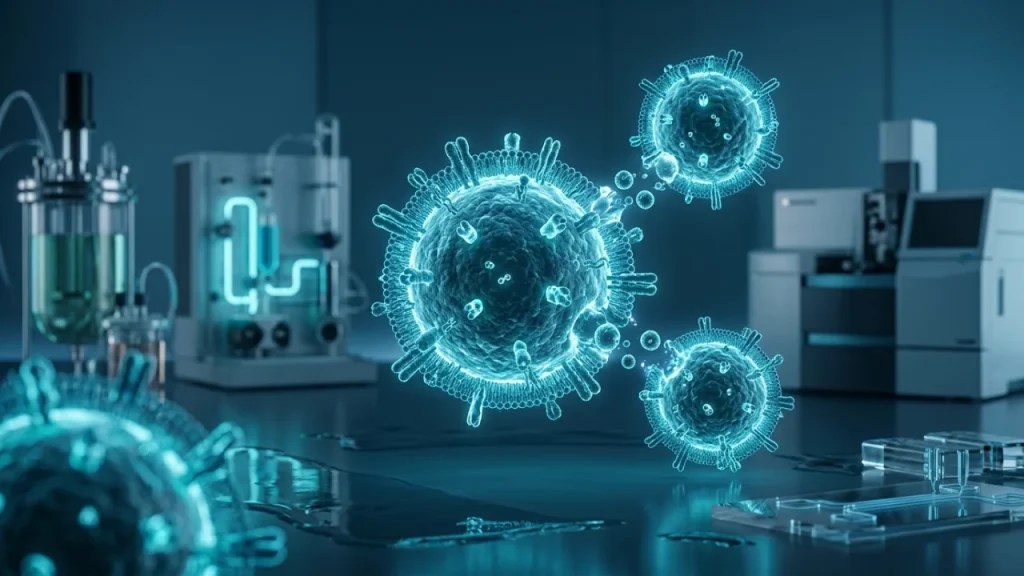
What Are Exosomes and Why Tampa Matters
Understanding Exosomes: Nature’s Tiny Messengers
Imagine your body’s cells are like a vast network of cities. They need to communicate. They don’t use phones or emails. Instead, they send tiny biological packages. These packages are called exosomes.
Exosomes are incredibly small vesicles. Think of a vesicle as a microscopic bubble. It is made from the same material as a cell’s outer membrane. Billions of these natural messengers travel through your bloodstream right now. They carry vital cargo from one cell to another.
What’s inside these tiny packages? The cargo is precise and powerful. It contains instructions and tools. – Genetic blueprints like RNA. – Proteins that can change a cell’s behavior. – Signaling molecules that give orders.
A liver cell can send an exosome to a muscle cell. The message might be about managing energy. A stem cell can dispatch exosomes to damaged tissue. These messengers can tell the damaged cells to repair themselves. This is a core reason scientists are so excited. Exosomes offer a natural way for the body to heal.
Disease changes this communication. For example, cancer cells are prolific senders. They can release ten times more exosomes than healthy cells. Their messages are dangerous. They might tell tumors to grow new blood vessels. They could convince the immune system to stand down. Studying these bad messages helps us understand how cancer spreads.
The potential for medicine is vast. Researchers see exosomes as next-generation delivery vehicles. Because they are natural, the body may not reject them. Scientists could load them with therapeutic cargo. This cargo could target a specific problem.
Think of a damaged heart after a heart attack. Exosomes could be designed to go only to those heart cells. They would deliver instructions to reduce scarring and promote healing. The same idea applies to brain diseases, arthritis, and wound repair. The goal is to use the body’s own system with enhanced precision.
This fundamental science is happening globally. It requires special labs and collaboration. This brings us to the growing work in exosomes Tampa. The city has become a key center for this research. Its scientific environment supports the complex work needed to understand these tiny messengers.
The journey from basic biology to new treatments is long. Understanding exosomes is the essential first step. They are not just cellular debris. They are a sophisticated communication network. Unlocking their code could change how we treat many diseases.
Why Tampa Became an Exosome Research Center
Tampa’s rise in exosome science is no accident. It resulted from deliberate planning and unique local strengths. The city built an ecosystem where discovery thrives. This ecosystem has several key parts.
First, Tampa benefits from major research institutions. These places have deep expertise in health and disease. They attract top scientists from around the world. These researchers need advanced tools to study tiny vesicles. Tampa’s labs invested in that equipment early.
Special microscopes are essential. They let scientists see exosomes directly. Other machines can analyze thousands of proteins at once. This technology is expensive and complex. Concentrating it in one geographic area creates efficiency. Scientists can share resources and ideas easily.
Collaboration is the second major factor. Breakthroughs rarely happen in isolation. Tampa’s research culture encourages teamwork across fields. A biologist might work with a bioengineer. A doctor treating patients partners with a lab scientist. This mix of skills speeds up progress.
Exosome research needs this mix. Understanding the basic biology is just one step. Turning that into a treatment requires engineering and clinical knowledge. Tampa’s network connects all these dots.
The third factor involves a critical local resource. The region has a strong focus on specific medical fields. These include cancer research, heart disease, and brain disorders. These are exactly the areas where exosome therapy shows great promise.
Scientists here study exosomes in real-world contexts. They ask specific questions. How do exosomes from a brain tumor help it spread? Can we intercept those messages? This disease-focused approach gives the research clear direction and purpose.
Funding and support form the fourth pillar. Long-term scientific work needs steady investment. Both public grants and private backing have flowed into exosomes Tampa projects. This financial stability allows researchers to pursue big, long-term questions.
They can run multi-year studies. They can take risks on innovative ideas. Consistent funding turns a short project into a sustained research program.
Finally, Tampa has a talent pipeline. Local universities train the next generation of scientists. Students learn about exosome biology in their classrooms. They then get hands-on experience in nearby labs. This creates a continuous flow of skilled researchers into the field.
All these elements combine to create a powerful center of gravity. – World-class institutions provide the anchor. – A collaborative culture breaks down barriers. – Specialized medical expertise focuses the work. – Strategic funding fuels long-term projects. – Educational programs renew the talent pool.
This environment tackles the big challenges in exosome science. One challenge is manufacturing. How do you produce pure exosomes at a large scale? Another is targeting. How do you ensure exosomes go only to sick cells? Tampa teams are working on these problems right now.
The city’s role matters for future medicine. Discoveries made here will influence global health. As the field moves toward clinical trials, Tampa’s integrated model is a blueprint. It shows how to translate basic science into real-world benefits.
The work continues to build on this strong foundation. The next phase involves turning these research insights into reliable, standardized tools for doctors and patients everywhere.
How Exosomes Tampa Drives Medical Innovation
Exosomes carry molecular messages that can change how cells behave. This makes them powerful tools for medicine. Researchers in Tampa are turning this basic science into new kinds of treatments. Their work tackles diseases in new ways.
One major area is regenerative medicine. Damaged tissues often cannot heal themselves. Scientists can load exosomes with specific instructions. These instructions tell the body’s own cells to start repair work. For example, exosomes can carry signals that reduce inflammation. They can also promote the growth of new blood vessels. This approach helps injured tissues recover faster. It is being studied for heart damage after attacks. It is also used for chronic wounds that will not close.
Another key focus is targeted drug delivery. Many powerful drugs hurt healthy cells along with sick ones. Tampa teams are designing exosomes as precise delivery vehicles. They modify the outer surface of these tiny vesicles. The modifications act like a postal code. They guide the exosome directly to a diseased organ or even a tumor. Once there, the exosome releases its cargo. This cargo could be a traditional chemotherapy drug. It could also be a new genetic therapy. This method aims to make treatments stronger and safer.
The diagnostic potential is equally important. Exosomes travel through all body fluids like blood. They carry a snapshot of the cells they came from. A tumor cell sheds exosomes that are different from healthy ones. Scientists in Tampa are developing sensitive tools to catch these differences. A simple blood test could one day find cancer very early. It could also show if a treatment is working without needing surgery.
The work on exosomes Tampa generates progress in three clear steps: – First, researchers at local institutions discover a new biological signal. – Next, they engineer exosomes to carry or target that signal. – Finally, collaborative teams test these engineered exosomes in models of disease.
This pipeline turns observations into potential solutions.
Beyond specific diseases, this innovation changes how we think about therapy. Exosomes are natural biological particles. The body may accept them better than synthetic materials. They can cross barriers that often block drugs, like the blood-brain barrier. This opens doors to treating brain conditions. Tampa’s integrated environment speeds up testing of these ideas.
The drive for medical innovation here solves practical problems. A big problem is how to store and ship exosome-based therapies without them breaking down. Tampa labs are creating stable forms that could be used in clinics everywhere. Another problem is making sure every batch is identical and pure. Standardized manufacturing methods are a key part of the research.
All this work points toward a future of more personal medicine. Doctors might use exosomes from your own body to aid your healing. Or they might use engineered ones to deliver treatment exactly where you need it. The ongoing research in Tampa moves these ideas from the lab toward the patient’s bedside. It shows why concentrating expertise in one collaborative hub matters for global health advances.
The next questions involve bringing these innovations safely to people through careful clinical trials.
The Economic Impact of Exosome Research in Tampa
Exosome research in Tampa creates high-value jobs. These are not just lab positions. The work requires skilled technicians, clinical trial managers, and manufacturing specialists. It also needs people in regulatory affairs, quality control, and business development. This demand strengthens the local workforce. It attracts talent from across the country. Each new research grant or company startup adds more positions. This growth is steady and knowledge-based.
The sector attracts significant investment. Venture capital firms fund promising biotech startups. Federal grants from agencies like the NIH support academic projects. This incoming money flows through the local economy. It pays salaries. It funds lab supplies from local vendors. It supports construction for new facilities. This investment cycle makes the biomedical field a powerful economic engine. It diversifies Tampa’s economy beyond tourism and traditional industries.
New companies are forming regularly. Many spin out from university discoveries. These startups focus on specific applications of exosome technology. Some work on diagnostics. Others develop therapies. This entrepreneurial activity is a direct result of the collaborative environment. A successful startup can grow rapidly. It may lease larger office or lab space. It will hire more employees. This commercial activity clusters around research institutions, creating a visible innovation district.
Supporting industries also benefit. Specialized services grow to meet researchers’ needs. This includes legal firms expert in patents and intellectual property. It includes marketing agencies for science communication. Companies that provide advanced laboratory equipment see more sales. Even shipping and logistics firms adapt to handle sensitive biological materials. This creates a wider network of economic activity linked to exosomes Tampa researchers study.
The focus on manufacturing is particularly important for long-term stability. Moving from lab-scale to large-scale production is a major challenge. Solving it requires engineering expertise and new facilities. Establishing local manufacturing plants would be a significant economic win. It creates durable industrial jobs. It positions the region as a production hub, not just a research center. This ensures the economic benefits last beyond initial discovery.
Property and infrastructure see direct impacts. Demand for modern laboratory space is rising. This encourages developers to build or retrofit buildings in specific areas. These projects increase property values. They also lead to improvements in local infrastructure to support advanced research facilities. The city invests in becoming an even more attractive place for this science.
The economic impact builds on itself. Success stories attract more attention. More attention draws more funding and more companies. This creates a virtuous cycle of investment and innovation. A strong local economy in this sector then funds further research. It allows for riskier, pioneering studies that could lead to bigger breakthroughs.
Training and education programs expand to meet industry needs. Local colleges and universities develop specialized courses. They create certificate programs for technical roles. This ensures a pipeline of local talent is ready for these new jobs. It keeps skilled graduates in the Tampa area. This educational component secures the region’s future as a leader in the field.
Ultimately, the economic story is about building lasting capacity. The goal is not just a temporary boom. It is to establish a resilient, knowledge-driven sector that provides careers and prosperity for decades. The work on exosomes today lays that foundation.
This financial growth enables the next critical phase: rigorous clinical trials to ensure safety and efficacy for patients everywhere
How Exosomes Work in the Human Body
How Cells Create and Release Exosomes
Cells create exosomes through a careful, multi-step process. It starts inside the cell. A part of the cell membrane folds inward. It forms a small pouch called an early endosome. This pouch captures various molecules from inside the cell. These molecules include proteins and genetic material like RNA.
The early endosome then changes. It matures into a late endosome. During this change, the membrane of the endosome buds inward again. It creates many tiny vesicles inside the larger structure. This creates a multi-vesicular body, or MVB. Think of an MVB like a microscopic shipping container. The tiny vesicles inside are the individual packages ready for export.
Each tiny vesicle inside the MVB is a future exosome. The cell carefully loads these vesicles with specific cargo. This cargo is not random waste. It is a selected mix of signaling molecules. The cargo reflects the cell’s current state and needs. A stressed cell sends different signals than a healthy one.
The cell must then decide what to do with this MVB. It has two main choices. The MVB can fuse with a structure called a lysosome. Lysosomes are the cell’s recycling centers. They break down the contents for reuse. Alternatively, the MVB can travel to the cell’s outer membrane.
For exosome release, the MVB takes the second path. It moves through the cell’s internal transport system. It reaches the inner surface of the cell membrane. The membrane of the MVB then fuses with the cell’s own outer membrane. This fusion opens the MVB to the outside world.
The tiny vesicles are now released into the extracellular space. They are now called exosomes. This release is an active process for communication. It is not just cellular garbage disposal.
The entire sequence has several key steps: – Invagination of the cell membrane to form an early endosome. – Maturation into a late endosome and inward budding to create an MVB. – Selective loading of molecular cargo into the internal vesicles. – Transport of the MVB to the cell periphery. – Fusion with the plasma membrane and release of exosomes.
Different cell types release exosomes at different rates. An immune cell, for instance, might use them to alert other cells to an infection. A neuron might use them to support nearby brain cells. The released exosomes enter bodily fluids. They travel in blood, saliva, and spinal fluid.
Their small size is crucial for this journey. Most exosomes are between 30 and 150 nanometers in diameter. This is about one thousandth the width of a human hair. Their tiny size lets them move easily through tissues and circulation.
Research in places like Tampa has helped map these details. Scientists use advanced microscopes to watch parts of this process. They also use biochemical tests to analyze the exosome cargo. Understanding how cells create and release exosomes is the first step. The next logical question is what happens after they are released. How do they find and affect another target cell? This delivery mechanism is just as precise as their formation.
What Exosomes Carry Inside Their Protective Shells
Exosomes carry a precise molecular package from their parent cell. This cargo is not random. It is carefully selected during the vesicle’s formation inside the cell. Think of an exosome as a tiny shipping container. Its contents are the valuable delivery.
The protective lipid shell keeps the cargo safe during travel. This is crucial for moving fragile molecules through harsh environments like the bloodstream. Inside, exosomes transport three main types of biological material.
First, they carry proteins. These can be enzymes to start chemical reactions in a target cell. They can also be signaling proteins that deliver instructions. Some proteins on the exosome’s outer surface act like address labels. They help the vesicle find the right cell to deliver its package.
Second, exosomes transport lipids. These are not just parts of the shell. Specific lipids inside can be used for energy by the receiving cell. Other lipids act as signals themselves. They can influence inflammation or help repair cellular membranes.
Third, and perhaps most importantly, exosomes carry nucleic acids. This includes RNA molecules. Messenger RNA (mRNA) can provide blueprints for making new proteins. MicroRNA (miRNA) acts as a control switch. It can turn specific genes in the target cell on or off.
The type of cargo defines the exosome’s mission. An exosome from a stem cell might carry growth factors and RNA to promote healing. An exosome from a cancer cell might carry signals that help tumors grow. Researchers in exosomes Tampa studies analyze this cargo to understand disease.
Scientists can open these vesicles in the lab to see what is inside. They use techniques like mass spectrometry for proteins. They use sequencing machines to read the RNA codes. This cataloging reveals health and disease states.
For example, exosomes from a healthy heart muscle cell carry different miRNAs than those from a stressed heart. This difference makes exosomes powerful biomarkers. Doctors could one day use a simple blood test to check exosome cargo for early disease signs.
The cargo is also the basis for potential therapies. Scientists can load exosomes with therapeutic molecules. They might pack them with a specific miRNA to correct a genetic problem. Or they could fill them with a drug that needs protection until it reaches its target.
The diversity of cargo is vast. A single exosome may hold hundreds of different protein molecules. It may contain thousands of RNA fragments. This complexity allows for sophisticated communication between cells.
Understanding this cargo is key to the promise of exosome science. It explains how a tiny vesicle can change a cell’s behavior without direct contact. The next step is seeing how this loaded container delivers its instructions to a target cell’s machinery.
How Exosomes Travel Between Different Cells
Exosomes begin their journey inside a cell. The cell creates them in a special compartment called an endosome. This endosome forms smaller vesicles inside itself. These internal vesicles are the future exosomes. The cell then releases this entire package to the outside.
The cell pushes this loaded endosome to its outer membrane. The two membranes fuse together. This process opens the endosome to the extracellular space. The tiny vesicles inside are now released. They are now free exosomes swimming in the body’s fluids.
These fluids act as a transportation network. Exosomes travel through blood plasma. They move within lymphatic fluid. They navigate the cerebrospinal fluid around the brain and spine. This system allows them to reach distant parts of the body.
Their movement is not random. Exosomes have targeting signals on their surface. These signals act like molecular postal codes. They help the vesicle find the right cell type. A liver cell exosome often seeks another liver cell.
The signals are often proteins or sugars. They stick out from the exosome’s lipid membrane. These molecules can bind to matching receptors on a target cell’s surface. This binding is like a key fitting into a lock.
The journey faces many obstacles. The bloodstream has immune cells that clear debris. Enzymes in fluids can break down molecules. Physical forces like shear stress could rupture the vesicles. The exosome’s durable membrane protects its cargo from these threats.
Distance is another major challenge. The body is vast at a cellular scale. Exosomes use the body’s existing flow systems to cover long distances. They are passive passengers in the blood’s current. Their surface signals ensure they get off at the right stop.
Once an exosome finds its target cell, several things can happen. The most common is direct fusion. The exosome membrane merges with the cell’s membrane. This merger dumps the exosome’s cargo directly into the cell’s interior.
Another method is endocytosis. The target cell engulfs the exosome. It wraps part of its membrane around the vesicle. It then pulls it inside in a new bubble. The cell then breaks this bubble down to access the cargo.
Sometimes, exosomes only deliver a surface signal. They bind to a receptor on the target cell without entering it. This binding alone can trigger a change inside the cell. It acts like a switch being flipped.
The efficiency of this delivery is remarkable. Studies show a significant portion of released exosomes reach specific targets. This precision is why they are such effective communicators. Their natural design optimizes for successful delivery.
The entire travel process has three main phases. – Release from the parent cell into bodily fluids. – Transport and navigation through those fluids. – Binding and delivery to the target cell.
Each phase must work perfectly for the message to get through. A failure in navigation means the exosome is lost. A failure in binding means the message is ignored. The system’s reliability is key to its function in health.
Cancer cells exploit this system. They send out many more exosomes than healthy cells. These tumor exosomes travel to other organs. They can prepare those organs for cancer spread. This shows how powerful this travel network can be.
Researchers in exosomes Tampa projects study how to hijack this travel system. They want to design synthetic exosomes that follow these natural routes. The goal is to create targeted drug delivery vehicles that use the body’s own pathways.
Understanding this journey completes the picture of exosome function. We now see how a message is written, packaged, sent, and received. This knowledge allows scientists to intercept faulty messages or send new therapeutic ones through this biological postal service.
Why Healthy Cells Use Exosomes to Communicate
Healthy cells constantly send exosomes to maintain order. This communication is vital for life. Think of your body as a vast city. Cells are the individual citizens and buildings. Exosomes are the messages they send to coordinate everything. Without this chatter, systems would fail.
Cells use exosomes for several key jobs. One major job is waste removal. Cells package old or broken proteins into exosomes. They then ship this material out for disposal. This keeps the cell clean and functioning well.
Another critical job is immune system coordination. For example, when a cell detects a virus, it can send out exosomes. These vesicles carry warning signals. They alert nearby cells to boost their defenses. They also inform immune cells about the threat’s location. This creates a rapid, organized response to infection.
Exosomes also help with tissue repair. After an injury, stem cells release special exosomes. These vesicles travel to the damaged site. They deliver instructions that tell local cells to grow and heal. They reduce inflammation. This helps the body fix itself efficiently.
Development and growth rely on exosomes too. In a growing embryo, cells use exosomes to guide each other. They tell neighboring cells what type to become. They help form organs and limbs correctly. This precise messaging ensures proper development from the start.
The content of a healthy exosome is carefully chosen. It typically includes: – MicroRNAs that regulate gene activity in the target cell. – Growth factors that encourage cell survival. – Enzymes that help with local chemical reactions. – Signal proteins that trigger specific pathways.
This cargo is not random. The parent cell selects it based on current needs and conditions.
Research into exosomes Tampa often highlights this normal function. Scientists study how healthy communication works. They need this baseline to spot when things go wrong. Understanding these natural roles is the first step toward medical applications.
Exosome communication is a two-way street. Cells do not just send messages. They also receive them. A cell constantly adjusts its behavior based on the exosomes it gets. This creates a dynamic, responsive network throughout your body.
This system is incredibly efficient. It uses the body’s own fluids as a highway. It does not require external energy beyond normal metabolism. The messages are protected in their lipid bubble. This ensures they arrive intact at their destination.
The volume of this traffic is astounding. Billions of exosomes move through your bloodstream right now. They are in your saliva and other fluids too. This constant flow of information is a sign of health. It shows your cells are talking and cooperating as they should.
Problems start when this communication breaks down. If exosome signals become confused, disease can follow. Chronic inflammation can disrupt normal messaging. Aging can change the quality of exosome cargo.
Scientists are fascinated by this natural technology. They see how evolution perfected this delivery system over millions of years. Now, they aim to learn from it. By copying these healthy processes, they hope to create new therapies.
The study of normal exosome function provides a powerful blueprint. It shows us how the body maintains balance and health through constant, microscopic conversation. This foundational knowledge directly informs the next logical question: what happens when this cellular communication goes wrong?
Tampa’s Exosome Research Facilities and Infrastructure
State-of-the-Art Labs for Exosome Studies in Tampa
Tampa hosts several major research centers dedicated to extracellular vesicle science. These facilities form the core of the city’s biomedical ecosystem. They provide the advanced tools needed for exosome work. This infrastructure attracts scientists from across the globe.
The labs specialize in isolating and studying these tiny particles. This is a major technical challenge. Exosomes are one hundred times smaller than a typical cell. Researchers cannot simply see them under a regular microscope. Specialized equipment is required to handle them.
A key piece of technology is the ultracentrifuge. This machine spins samples at incredible speeds. The force separates exosomes from other components in blood or cell culture. The process is gentle but precise. It preserves the exosomes’ delicate structure and cargo.
Many labs also use nanoparticle tracking analysis. This technology uses laser light to visualize exosomes. It lets scientists count particles and measure their size. They can see if a sample contains millions of exosomes or just a few thousand. This data is crucial for quality control.
Flow cytometry is another essential tool. It can sort exosomes based on surface markers. Think of it as a high-speed mail sorter for microscopic vesicles. It identifies exosomes from different cell types. This helps trace a vesicle’s origin and potential function.
The work requires extremely clean environments. Contamination can ruin sensitive experiments. Many labs have dedicated clean rooms. Scientists wear gowns, gloves, and hair nets. The air is filtered constantly. This prevents dust or microbes from interfering.
Cell culture facilities are equally important. Scientists grow human cells in controlled conditions. They study how these cells produce and release exosomes. They can change the cell’s environment to see how it affects the vesicles. This reveals the triggers for healthy or diseased messaging.
Advanced imaging cores offer a closer look. Electron microscopes can take pictures of individual exosomes. These images show the classic cup-shaped structure of the vesicles. Scientists can see the lipid bilayer that protects the molecular message inside.
Biobanks are a critical part of the infrastructure. These are secure, frozen libraries of biological samples. They store blood, tissue, and isolated exosomes from donors and patients. This preserved material allows for long-term studies and comparisons.
The scale of operation is significant. A single research center may process hundreds of samples weekly. Teams often work around the clock. Experiments can run for days or weeks without stopping. The goal is to generate reliable, reproducible data.
Collaboration is built into the design of these spaces. Open lab layouts encourage conversation between researchers. Shared equipment cores are common. A scientist studying cancer exosomes works near one focused on neurodegenerative disease. This proximity sparks new ideas.
Supporting this work requires robust computational power. The data generated is massive. Bioinformatics teams analyze complex genetic and protein information from exosome cargo. They look for patterns linked to health or disease states.
This concentration of technology creates a powerful synergy for discovery in exosomes Tampa. Researchers have the tools to ask detailed questions about cellular communication. They can move quickly from an idea to an experiment. This accelerates the entire field.
The presence of these facilities directly impacts the local economy and talent pool. It creates high-skill jobs in science and engineering. It also trains the next generation of researchers through university partnerships.
This advanced infrastructure turns theoretical knowledge into practical insight. It allows scientists to decode the exact messages within exosomes. The next step is applying this knowledge to develop new diagnostic tools and therapeutic strategies, a natural progression for Tampa’s research community.
Special Equipment for Studying Exosomes Tampa Uses
Studying exosomes requires specialized tools. Scientists cannot use standard lab microscopes to see them easily. Exosomes are far too small. They are about one-thousandth the width of a human hair. This demands powerful technology.
The first major challenge is isolation. Exosomes must be separated from blood or cell culture fluid. This fluid contains many other particles. Scientists use several key methods for this clean separation.
Ultracentrifugation is a common workhorse. Machines spin samples at incredible speeds. These speeds can exceed 100,000 times the force of gravity. Heavier particles sink to the bottom first. Lighter exosomes form a pellet later. This method is reliable but can be slow.
Size-based chromatography is another vital tool. Scientists push the liquid through a column filled with tiny beads. The beads have precise pores. Larger molecules get trapped or slowed down. Smaller exosomes flow through more quickly. This separates them by physical size.
Precipitation kits use chemical solutions to change the liquid’s properties. Exosomes become less soluble. They fall out of solution gently. This method is often faster and needs less expensive equipment. Each isolation method has its own strengths for different research goals in exosomes Tampa.
After isolation, scientists need to confirm what they have. They must prove the particles are truly exosomes. Nanoscale flow cytometry is a crucial instrument here. It analyzes particles one by one as they flow past a laser.
The machine measures two key things. It checks the particle’s size. It also detects specific protein markers on the surface. True exosomes carry markers like CD63 and CD81. This double-check is essential for quality research.
Visual confirmation comes from electron microscopes. These are not normal microscopes. They use a beam of electrons instead of light. This provides much higher magnification. Scientists can see the classic cup-shaped structure of dried exosomes. It offers a direct visual proof.
The next step is cargo analysis. This is where the real messages are read. Scientists break open the isolated exosomes to see what’s inside. The cargo includes proteins, RNA, and lipids.
Mass spectrometers identify proteins. They weigh protein fragments with extreme accuracy. Each protein has a unique weight signature. The machine compares this to databases to name the protein.
For genetic material, sequencing platforms are key. They read the RNA sequences inside the exosomes. This shows what genetic instructions the exosome was carrying. Bioinformatics software then finds patterns in this complex data.
Advanced labs also use instruments for real-time tracking. Fluorescence labeling lets researchers tag exosomes with a glowing marker. They can then watch how these exosomes move and which cells they enter. This reveals communication pathways in living systems.
The equipment list for modern exosomes Tampa research is extensive. – Ultracentrifuges for isolation. – Nanoscale flow cytometers for verification. – Electron microscopes for imaging. – Mass spectrometers for protein cataloging. – Sequencers for RNA analysis.
This toolkit allows researchers to move from a mixed sample to detailed knowledge. They learn the exosome’s source, its cargo, and its potential function. This precise physical analysis forms the foundation for all future applications. Understanding these tools shows how abstract ideas become concrete, measurable science that can lead to new medical insights.
How Tampa Researchers Isolate Pure Exosomes
Isolating pure exosomes is like finding specific needles in a haystack. The haystack is a complex biological fluid like blood or cell culture medium. Researchers need to separate the tiny exosomes from everything else. They must do this without damaging the exosomes. Tampa laboratories use several trusted methods to achieve this purity.
One common technique is ultracentrifugation. This method uses very high spinning speeds. A sample is placed in a special tube. This tube goes into a machine called an ultracentrifuge. The machine spins it incredibly fast. This creates strong centrifugal forces. Different particles separate based on their size and density. Larger particles sink to the bottom first. Exosomes form a pellet at the bottom after a long spin. The liquid above is carefully removed. The exosome pellet is then washed and spun again. This process increases purity. It is a foundational method in many exosomes Tampa research projects.
Another popular method is size-exclusion chromatography. Here, a sample is passed through a column filled with porous beads. The beads act like a sieve. Large molecules cannot enter the pores. They flow through the column quickly. Smaller molecules enter the pores and move slowly. Exosomes are too big to enter most pores. They elute in the early fractions. This method is gentle. It keeps exosomes intact and functional.
Precipitation is a simpler, faster technique. A special polymer solution is added to the sample. This solution changes the solubility of the exosomes. It makes them less soluble in the liquid. The exosomes clump together and fall out of solution. They are then collected by a low-speed spin. This method is good for quick experiments. However, it can sometimes co-precipitate other non-exosome material.
Many modern labs now use immunoaffinity capture. This method targets exosomes by their surface markers. Antibodies are used like magnets. These antibodies stick to specific proteins on the exosome’s outer membrane. The antibodies are attached to magnetic beads or a column. When the sample flows past, only exosomes with the right markers are caught. Everything else washes away. This gives very pure exosomes from a specific cell type.
The choice of method depends on the research goal. – Ultracentrifugation is excellent for large yields. – Size-exclusion chromatography is ideal for preserving biological activity. – Immunoaffinity capture provides the highest specificity for certain exosome types.
After isolation, scientists must confirm what they have. They check the size and concentration of the particles. A tool called a nanoparticle tracking analyzer does this. It shines a laser through the sample and watches particles move. Software calculates their size and number. Researchers also check for classic exosome marker proteins through tests like Western blotting.
Each isolation method has trade-offs between purity, yield, speed, and cost. Tampa’s collaborative environment lets researchers choose the best tool for their question. Pure isolation is not just a technical step. It is the essential foundation for all reliable discovery. Clean samples ensure that any signal scientists find later truly comes from exosomes, not from contaminants. This rigorous first step supports the entire local ecosystem of exosomes Tampa innovation, leading directly to trustworthy results in diagnostics and therapy development
Storage and Handling of Exosomes in Tampa Labs
Once isolated, exosomes are fragile. Their biological activity can degrade quickly without proper care. Tampa labs treat these nanoscale vesicles with great precision. This ensures their research value remains intact from the lab bench to future experiments.
Storage temperature is the first critical decision. Most labs use ultra-cold freezers for long-term keeping. – -80°C freezers are the standard workhorses. They dramatically slow all chemical and physical decay. Exosomes can remain stable here for months or even years. – Liquid nitrogen offers even colder storage at -196°C. This is used for extremely valuable or irreplaceable samples. All biological activity essentially stops at this deep cold.
But freezing itself can damage the exosomes. Ice crystal formation can rip their delicate lipid membranes. Tampa researchers use special techniques to prevent this harm. They add protective substances called cryoprotectants to the exosome solution before freezing. These compounds, like sugars or specific proteins, shield the vesicles from ice damage.
How samples are packaged is equally vital. Researchers avoid large volumes in single containers. They instead use small, labeled tubes called cryovials. Scientists divide a sample into many small portions called aliquots. This prevents repeated freeze-thaw cycles of the main stock. Each small vial is used only once. This practice maintains consistency across long-term studies central to exosomes Tampa research projects.
The thawing process requires careful control. Rapid warming in a 37°C water bath is common. Gentle mixing ensures an even temperature. Scientists never let samples thaw at room temperature. They also avoid vortexing or harsh shaking, which can destroy exosome structure. Once thawed, the sample is typically kept on ice and used quickly for the next experiment.
Handling exosomes during experiments demands clean technique. Researchers use filtered pipette tips and sterile buffers. This prevents contamination from dust or microbes. They also track sample history meticulously in digital lab notebooks. Each entry notes isolation date, storage time, freeze-thaw count, and handling details.
Advanced Tampa facilities monitor their storage units constantly. Automated systems track freezer temperatures around the clock. Alerts go to scientists’ phones if a unit fails. Backup generators turn on during power outages. This protects years of precious research material.
The goal of all these steps is functional preservation. Scientists need more than just particles in a tube. They need vesicles that still carry their original biological messages. Proper storage keeps exosome membranes intact. It also protects the internal cargo of proteins and RNA.
Quality checks continue after storage. Researchers often re-test a thawed sample’s size and marker proteins. They compare this data to readings taken just after isolation. This confirms the handling protocol worked. Stability data from Tampa labs helps set global standards for exosome biobanking.
This meticulous attention to storage forms a reliable bridge. It connects the isolation process to downstream applications. Stable, well-characterized exosome samples fuel consistent research outcomes. They enable trustworthy diagnostics and reproducible therapy development. The city’s focus on robust infrastructure supports every stage of discovery, ensuring that valuable findings are built on a foundation of preserved integrity.
Exosome Applications in Regenerative Medicine
How Exosomes Help Repair Damaged Tissues
Exosomes act as natural repair messengers in the body. When tissue gets damaged, cells release these vesicles. They carry instructions to the site of injury. This starts a coordinated healing process. The cargo inside exosomes tells other cells what to do.
These tiny packages contain different types of information. They carry proteins that can signal growth. They also contain nucleic acids like RNA. This RNA can reprogram how a recipient cell behaves. The membrane protects this cargo during its journey. This is why proper storage in exosomes Tampa research is critical. Scientists must keep these messages intact for studies.
The healing process involves several key steps. Exosomes help manage each one.
- First, they reduce immediate inflammation. They send signals to calm the immune system. This prevents excessive damage from the body’s own response.
- Next, they promote new blood vessel growth. This step is called angiogenesis. New vessels bring oxygen and nutrients to the damaged area. This is vital for repair.
- Then, they stimulate cell proliferation. They encourage local cells to divide and multiply. This replaces cells that were lost or injured.
- Finally, they guide tissue remodeling. They help organize new cells into functional structures. This restores the tissue’s original job.
For example, consider muscle repair after a strain. Inflammatory cells rush to the injury. Local muscle stem cells become active. Exosomes from various cells coordinate this. They tell inflammatory cells when to stand down. They also direct stem cells to become new muscle fibers. This repairs the tear efficiently.
Cardiac tissue repair after a mild heart attack shows another path. Damaged heart cells release exosomes. These vesicles encourage new blood vessels to form around the scar. They also send survival signals to stressed heart cells nearby. This can limit the overall area of damage. It improves the heart’s recovery of function.
In nerve regeneration, the challenge is different. Nerves heal very slowly. Exosomes from support cells can create a better environment for growth. They clear away debris that blocks regrowth. They also provide a scaffold for nerve fibers to follow. This can potentially restore lost connections.
Skin wound healing is a visible example. Exosomes help skin cells migrate across a cut. They accelerate the closure of the wound. They also modulate collagen production. This helps minimize scarring during the final remodeling phase.
The source of the exosomes matters greatly for therapy. Different parent cells create vesicles with different instructions. Mesenchymal stem cell exosomes are common in regenerative studies. These cells naturally aid in tissue support and repair. Their exosomes mirror these abilities. Immune cell exosomes can finely tune inflammation. The choice depends on the target injury.
Researchers in Tampa and worldwide are mapping these precise signals. They ask which cargo components are most important for each repair step. Is it a specific microRNA? Is it a surface protein? Finding these answers allows for more targeted therapeutic designs. Scientists could then enrich exosomes for particular healing factors.
This therapeutic potential relies entirely on quality control. Exosomes must be functionally potent when they reach a patient. They cannot have degraded during storage or transport. The rigorous protocols developed for biobanking ensure this. Stable, well-preserved exosomes deliver consistent, predictable healing signals to damaged tissues, turning scientific discovery into reliable medical promise.
The next logical question is how these repaired tissues integrate into a living system, leading to studies on systemic health effects and long-term outcomes from exosome therapies.
Exosome Therapy for Joint and Muscle Recovery
Exosomes offer a promising strategy for healing damaged joints and muscles. This approach targets common, painful conditions. These include osteoarthritis and sports injuries. The goal is to reduce pain and restore function. It works by addressing the root causes of degeneration.
Joint diseases like osteoarthritis involve a cycle of damage. Inflammation breaks down protective cartilage. This cushion between bones wears thin. Pain and stiffness follow. Traditional treatments often manage symptoms alone. They may not repair the tissue. Exosome therapy aims to change this process.
Exosomes deliver precise instructions to cells in the joint. They carry signals that can calm the immune system. This reduces harmful inflammation. They also encourage chondrocytes to produce new matrix components. Chondrocytes are the cells that maintain cartilage. Furthermore, exosomes may help slow cartilage breakdown. They do this by modulating enzymes that degrade tissue.
For muscle recovery, the mechanism is equally direct. After a strain or tear, muscle fibers need to regenerate. Satellite cells are stem cells for muscle. They must activate and fuse to repair damage. Research shows certain exosomes can trigger this activation. They promote the growth of new blood vessels too. This improved blood flow brings more oxygen and nutrients to the injured site.
The potential benefits for patients are clear. – Reduced pain and swelling in joints. – Improved range of motion and mobility. – Possible delay or avoidance of joint replacement surgery. – Faster return to activity after muscle injuries. – Longer-lasting results by supporting natural healing.
The science behind this is advancing rapidly in centers like those in exosomes Tampa research networks. Scientists are identifying which exosome cargo works best. For joints, exosomes rich in anti-inflammatory microRNAs are key. For muscles, exosomes carrying growth factors like VEGF are important. VEGF stimulates blood vessel formation.
Treatment typically involves a precise injection into the affected area. The exosomes come from a controlled laboratory process. They are not taken directly from a donor at the point of care. This ensures safety and consistency. Once injected, the vesicles begin their signaling work immediately. They interact with local cells to shift the environment from destructive to regenerative.
Clinical observations report positive outcomes. Patients often experience decreased pain within weeks. Functional improvements may continue for months as repair progresses. The therapy leverages the body’s own communication system. It guides tissues toward a natural healing pathway.
This local application for musculoskeletal problems connects to broader health. Successful joint repair improves overall mobility. Better mobility supports heart health and metabolic function. The next frontier examines how these localized treatments influence whole-body wellness over time, opening a discussion on systemic effects and long-term vitality.
Skin Regeneration Using Exosome Technology
Skin is the body’s largest organ. It constantly repairs itself. Exosomes offer a powerful way to support this natural process. These tiny messengers carry instructions to skin cells. They tell cells to rebuild and renew.
The science is clear. Exosomes from certain cell types are packed with specific signals. For skin, these signals focus on key tasks. One major task is making collagen. Collagen is the protein that gives skin its strength and fullness. As we age, collagen production slows. Exosomes can restart it. They deliver microRNAs that turn on the fibroblast cells. Fibroblasts are the skin’s collagen factories.
Another critical task is healing wounds. This includes cuts, burns, and even acne scars. Exosomes speed up this repair. They reduce inflammation quickly. They also direct new blood vessel growth to the injured area. This brings more oxygen and nutrients. The result is faster closure of the wound with less scarring.
Research in places like the exosomes Tampa biomedical community explores these uses. Scientists study which exosome sources work best for different skin concerns. The goal is to create targeted treatments.
The potential applications are broad. Here are a few key areas: – Repairing sun-damaged skin by restoring healthy cell function. – Improving the appearance of fine lines and wrinkles through collagen renewal. – Accelerating recovery after procedures like laser therapy. – Reducing hyperpigmentation by regulating melanin production in skin cells. – Enhancing moisture retention by supporting the skin’s barrier.
Treatment methods are evolving. Topical creams containing exosomes are one approach. These aim to deliver vesicles directly to the upper layers of skin. More profound effects may come from injections. Precise injections can place exosomes deeper into the dermis. This targets the living layer where collagen is made.
The process is elegant. Applied exosomes merge with local skin cells. They release their cargo of proteins and genetic material. This cargo reprograms the cell’s activity. The cell shifts from a passive state to an active repair state. It is not just adding a temporary filler. It is encouraging the skin to heal itself from within.
Safety is a prime advantage. Exosomes act as signaling tools. They do not replicate like live cells. They carry instructions and then are cleared by the body. Their effect is through changing cell behavior, not by staying permanently.
Clinical observations are promising. Patients see improvements in skin texture and elasticity. The effects develop over weeks and months as new collagen forms. This aligns with the body’s natural biological timelines.
The connection to overall health is direct. Skin is a barrier against infection. Effective repair maintains this crucial shield. Healthy skin also supports immune function and temperature regulation.
This technology moves beyond surface-level care. It uses the body’s own communication system for deep regeneration. The success in skin therapy complements progress in joint and muscle healing. Together, they highlight a unified principle: guiding innate repair mechanisms offers a powerful path in medicine. The next logical step is to examine how these targeted treatments might influence aging and wellness at a whole-body level, considering the systemic signals exosomes may carry.
Cardiac Repair Through Exosome Signaling
Heart muscle cells, called cardiomyocytes, have a limited ability to regenerate after injury. A heart attack leaves behind scar tissue. This scar weakens the heart’s pumping power. Exosome signaling offers a strategy to change this outcome. These tiny vesicles carry instructions that can alter the damaged environment.
The process starts after a heart attack. Dying and stressed cells release exosomes. These exosomes contain specific cargo. This cargo includes microRNAs and proteins. They send signals to the surviving cells. The goal is to shift the tissue from a state of scarring to one of repair.
Exosomes target several key problems at once. They do not work on just one issue. Their multi-pronged approach is their strength. – They reduce harmful inflammation. Exosomes can signal immune cells to calm down. This prevents further collateral damage to healthy heart tissue. – They promote new blood vessel growth. This is called angiogenesis. New vessels bring oxygen and nutrients to the injured zone. This is vital for survival of any recovering cells. – They encourage heart muscle cell survival. Exosome signals can block cell death pathways. This helps protect the vulnerable border zone around the scar. – They may even stimulate existing cells to divide. This can lead to a small degree of true muscle regeneration.
Research shows these effects are measurable. In studies, animals treated with exosomes after a heart attack show better heart function. Their ejection fraction, a measure of pumping strength, improves. Their scar size is often smaller. The treated heart tissue also shows more signs of new blood vessel formation.
The source of the exosomes matters for cardiac repair. Scientists study exosomes from many cell types. Mesenchymal stem cell exosomes are a major focus. These cells are naturally involved in healing. Their exosomes carry a potent mix of regenerative signals. Using just the exosomes avoids risks linked to whole cell transplants.
Safety is again a key point for heart applications. Exosomes act as transient messengers. They deliver their instructions and are cleared. They do not form tumors or block blood vessels. This makes them a attractive tool for a sensitive organ like the heart.
The potential extends beyond emergency heart attack care. Exosome therapy could help in chronic heart failure. In this condition, the heart is constantly stressed and inflamed. Regular exosome signaling might protect the muscle over time. It could slow the disease’s progress and improve quality of life.
The work happening in exosomes Tampa research hubs is accelerating this field. Local scientists are refining ways to produce clinical-grade exosomes. They are designing methods to target them specifically to heart tissue. This targeting could increase the treatment’s effectiveness and lower the needed dose.
Cardiac repair demonstrates a powerful principle. The body’s own communication system can be harnessed for major organ healing. The logic is similar to skin repair but on a larger scale. The signals reduce damage, improve blood flow, and support cell survival.
This success with heart tissue opens clear questions. If exosomes can aid the heart, what about other vital organs? The brain and the liver also suffer from poor regeneration after injury. The next frontier is applying these lessons to neurological and metabolic conditions, exploring how these universal messengers might heal different systems.
Exosomes in Cancer Research and Treatment
How Cancer Cells Use Exosomes to Spread
Cancer cells send out far more exosomes than normal, healthy cells do. This flood of tiny vesicles is not random. It is a key part of how tumors grow and spread. The exosomes act as advance scouts and saboteurs for the cancer.
Think of a tumor as a harmful headquarters. It packs exosomes with specific instructions. These instructions are meant to change the environment around the cancer. The goal is to make the body’s own systems help the tumor survive.
One major job of these exosomes is to suppress the immune system. Immune cells are the body’s natural defense army. They patrol for and attack invaders like cancer. Tumor-derived exosomes can disarm these guards.
They do this in several direct ways. – They carry signals that tell immune cells to become inactive. – They can mark cancer cells as “self,” making them invisible to attack. – They can exhaust immune cells, draining their energy to fight.
This creates a shield around the growing tumor. The local defense network is shut down. This allows the cancer to expand without interference.
Exosomes also help build a support network for the tumor. This is called preparing the “pre-metastatic niche.” Metastasis is when cancer spreads to new organs. It is the most dangerous stage of the disease.
A tumor in the breast or lung, for example, cannot just jump to the brain or liver. The new site must be ready to receive it. Cancer exosomes travel through the bloodstream to distant organs. They arrive long before any cancer cells get there.
These exosomes start making changes in the distant tissue. They encourage inflammation. They remodel local structures to make them more welcoming. They essentially lay out a welcome mat for traveling cancer cells later on.
The vesicles also break down natural barriers. Our tissues have strong, wall-like structures called basement membranes. These membranes keep cells in their proper place. Exosomes from tumors can carry enzymes that chew holes in these walls.
This creates an escape route for cancer cells. It is like the exosomes cut a hidden door in a fence. Then the tumor cells can slip out into the bloodstream or lymphatic system.
Communication with normal cells is another tactic. Cancer exosomes can fuse with healthy cells in other parts of the body. They deliver their cargo of proteins and genetic material. This can turn these normal cells into helpers.
For instance, they can instruct normal cells to build new blood vessels. Tumors need a constant supply of oxygen and food to grow. These new vessels provide it directly to the cancer. This process is called angiogenesis.
The genetic material inside exosomes is especially powerful. It often includes microRNAs. These are small pieces of genetic code that can control a cell’s behavior. A cancer’s microRNA can reprogram a distant cell’s functions.
It can tell that cell to grow faster. It can tell it to move toward the tumor. This turns healthy tissue into a partner for disease progression.
Research into exosomes Tampa labs focuses heavily on this dark side of exosome biology. Scientists are mapping exactly which cargo items cause which effects. They are looking for the most dangerous signals.
This knowledge is critical for two reasons. First, it helps us understand metastasis better. Stopping this spread is the main goal in curing many cancers. Second, it reveals new targets for treatment.
If we can block tumor exosomes from being made, we might slow the cancer. If we can intercept their messages, we could break its communication lines. We could also use these exosomes as early warning signs.
Because they are abundant and travel in blood, they make excellent biomarkers. Doctors could test a simple blood sample. Finding certain cancer exosomes could reveal an invisible tumor long before it shows up on a scan.
This paints a clear picture. Exosomes are a fundamental tool in the cancer toolkit. Their natural role in cell talk is hijacked for a harmful purpose. The same system that can heal a heart can also help a tumor thrive.
This duality makes the science complex but full of opportunity. Understanding how cancer uses these vesicles teaches us how to stop it. It also shows us how to better use exosomes for good, ensuring our therapies outsmart the disease’s own strategies. The next step is turning this enemy tactic into a weakness we can attack.
Detecting Cancer Early Through Exosome Analysis
Cancer cells send out far more exosomes than normal cells. This flood of vesicles creates a clear signal in a patient’s blood. Scientists can now find this signal long before a tumor is large enough to see.
The process starts with a simple blood draw. This liquid biopsy is much easier than a tissue biopsy. Researchers then separate the tiny exosomes from other blood components. They use special filters or centrifuges to do this.
Once isolated, the real detective work begins. Scientists look at two main things. First, they analyze the exosome surface. Cancer exosomes often have unique protein markers. These markers act like a return address, showing which cell sent them.
Second, they look inside the cargo. They check for genetic material like microRNA or DNA fragments. These pieces can carry the specific mutations of the tumor. Finding them is like finding a fingerprint at a crime scene.
This method offers several powerful advantages over traditional scans.
- It is minimally invasive. A blood test is simple and low-risk.
- It can detect cancer earlier. Tumors release exosomes when they are very small.
- It may show if treatment is working. Falling exosome levels could mean therapy is succeeding.
- It can help monitor for recurrence. Regular blood tests might catch returning cancer fast.
Research in exosomes Tampa and other hubs is making this process better. Teams are creating more sensitive tools to catch these vesicles. They are building detailed libraries of cancer signals. This helps match an exosome to a specific cancer type.
For example, pancreatic cancer is often found too late. Its exosomes, however, appear in blood early. A reliable test could save many lives. Similar work is ongoing for lung, breast, and ovarian cancers.
The goal is a routine screening test. Imagine a yearly physical that includes an exosome scan. This could catch dangerous cancers when they are most treatable. It turns the body’s communication system into an early warning network.
Of course, challenges remain. Not every exosome in blood is from cancer. Researchers must be sure their signals are correct. They must avoid false alarms that cause unnecessary worry. The technology must also become affordable for widespread use.
Yet the path forward is clear. By decoding the messages in these tiny bubbles, medicine gains a new sense. It can hear what the body’s cells are saying long before a tumor makes itself known. This shifts the fight from late-stage battles to early interventions.
Early detection changes everything for patient survival. The next logical step is using this knowledge not just to find cancer, but to fight it directly with targeted therapies.
Targeted Drug Delivery Using Engineered Exosomes
Cancer cells send out many exosomes. These tiny bubbles can travel far in the body. Scientists have a powerful idea. They can turn these bubbles into tiny drug trucks.
The goal is simple. Load a cancer drug into an exosome. Then send it directly to the tumor. This method is called targeted drug delivery. It is much smarter than standard chemotherapy.
Standard chemo floods the entire body. It hits healthy cells and causes harsh side effects. Targeted exosomes aim to change that. They seek out only the cancer cells. This spares healthy tissue.
How does this targeting work? Researchers use a natural trick. Exosomes have addresses on their surface. These are called targeting ligands. Think of them as GPS coordinates.
Scientists can engineer these addresses. They change them to match a specific tumor. For example, a breast cancer tumor might have a unique surface marker. An exosome can be designed to find that marker.
The process has several key steps. First, scientists choose a source for the exosomes. Often, they use human cells grown in a lab. These cells are like factories.
Next, they engineer those factory cells. They give them new instructions. The cells are told to make exosomes with special addresses and to pack them with medicine.
Then, the cells release the custom exosomes. Researchers collect and purify them. Finally, the exosomes are ready for delivery.
What kind of drugs can they carry? The list is growing. – Traditional chemotherapy drugs. – Newer genetic medicines like siRNA or miRNA. – Special proteins that can kill cancer cells.
This approach solves big problems. Some cancer drugs are very toxic. Packaging them in exosomes can hide them from the immune system. This prevents a bad reaction.
Other drugs are fragile. They break down in the bloodstream before reaching the tumor. The exosome’s lipid bubble acts as a protective shell.
Research in exosomes Tampa and other centers is testing this in labs. One promising area is brain tumors. The brain has a protective barrier. This barrier blocks most medicines.
Exosomes, however, can sometimes cross this barrier. Engineered versions could carry drugs straight to a brain tumor. This was very hard to do before.
Another challenge is drug resistance. Sometimes cancer cells pump medicine back out. Exosomes can bypass these pumps. They fuse with the cancer cell and dump their cargo inside.
The future looks like this: A patient is diagnosed with cancer through an exosome blood test. Doctors then analyze the tumor’s specific markers. They create a personalized batch of drug-carrying exosomes.
These exosomes would be given to the patient through an IV drip. They would travel through the bloodstream, hunting for the cancer. They would release their poison only where it is needed.
This reduces side effects like nausea and hair loss. It could allow for higher, more effective drug doses at the tumor site.
Of course, this science is still developing. Making these exosomes is complex and costly. Researchers must ensure they are safe and consistently effective.
Yet the principle is solid and inspiring. We are learning to hijack the body’s own communication system. We turn a tool used by cancer into a weapon against it.
This moves us beyond detection into precise intervention. The next question is how to boost the body’s own defenses using similar technology.
Stopping Tumor Growth with Exosome Therapies
Cancer cells use exosomes as tools for their own survival. They send out far more exosomes than healthy cells do. These tiny packets carry specific orders. The orders tell surrounding tissues to help the tumor grow.
One major command is: “Build new blood vessels.” Tumors need a constant supply of food and oxygen. This process is called angiogenesis. Exosomes from cancer cells can trigger it. They deliver signals that encourage blood vessel growth directly into the tumor.
New therapies aim to stop this command. Scientists are designing “decoy” exosomes. These decoys would bind to the signals. They would block the message from being received. Without new blood vessels, the tumor’s growth slows dramatically. It can even begin to shrink from starvation.
Exosomes also help cancer spread to other organs. This is called metastasis. It is the most dangerous stage of the disease. Before a cancer cell moves, it sends exosomes ahead. These exosomes act like scouts preparing a new site.
The scout exosomes land in distant organs like the liver or lungs. They make the environment there welcoming for cancer. They suppress local immune defenses. They also remodel the tissue structure. This creates a “pre-metastatic niche.” It is a landing pad for traveling cancer cells.
Stopping these scouts is a key research goal in exosomes Tampa studies and beyond. One approach uses engineered exosomes that carry different messages. These therapeutic exosomes could tell the distant organ to stay hostile to cancer. They could activate immune cells in that area instead of calming them down.
Another strategy targets the tumor’s communication network directly. Researchers are creating exosomes that bind to receptors on cancer cells. This binding can trigger a self-destruct sequence inside the cell. The process is known as apoptosis. It is a natural form of cell death that cancer normally avoids.
These therapeutic exosomes can also carry special RNA molecules. The RNA can silence critical genes inside the cancer cell. For example, it can turn off a gene that makes the cell immortal. Without that gene, the cancer cell ages and dies like a normal cell would.
The immune system is a powerful ally in this fight. But cancer often uses exosomes to hide from immune attack. Tumor-derived exosomes can wear disguises. They display proteins that tell immune cells, “I am friendly, do not attack.”
New treatments seek to remove this disguise. Some experimental exosome therapies act as warning flags. They attach markers to the cancer cells that shout “Danger!” to the immune system. This helps natural killer cells and T-cells find and destroy the tumor.
Combining these approaches could be very effective. A multi-step treatment might look like this: – First, deploy exosomes that cut off the tumor’s blood supply. – Second, send exosomes that block communication to distant sites. – Third, use exosomes that mark cancer cells for immune destruction. – Finally, use exosomes to deliver a final toxic drug payload directly inside weakened cells.
This layered attack tackles cancer on several fronts at once. It uses the body’s own communication system against the disease. The goal is to overwhelm the tumor’s defenses through smart biological tactics.
Of course, turning off growth signals must be very precise. Healthy cells also need to communicate for normal tissue repair. Scientists are working on targeting systems with extreme accuracy. They use unique markers found only on cancer cell exosomes as guides.
The progress in this area turns a major problem into a solution. Cancer’s aggressive use of exosomes reveals its weaknesses. Researchers are now learning to intercept and rewrite its messages. This moves therapy from blunt force to strategic interference.
The next logical step is enhancing natural healing using these same principles. If exosomes can carry harmful messages, they can also carry helpful ones for recovery after treatment.
The Future of Exosome Research in Tampa
Next-Generation Exosome Engineering Techniques
Scientists are now moving beyond using natural exosomes. They are learning to design them from the ground up. This field is called exosome engineering. It allows for extreme precision in medicine. Think of it as custom-building a microscopic delivery vehicle. Every part can be chosen for a specific job.
The first step often involves the exosome’s surface. This outer membrane is like the address label. Researchers can attach special molecules called targeting ligands. These ligands act like homing devices. They bind only to certain cell types. For example, a ligand might seek out only inflamed joint tissue or a specific cancer cell. This ensures the exosome’s cargo goes exactly where it is needed.
The cargo inside the exosome is equally important. Engineers can load exosomes with many different things. They are not limited to natural molecules. Potential cargo includes: – Synthetic drugs that normally cannot enter cells. – Specific RNA instructions to turn genes on or off. – Corrected DNA sequences to fix genetic errors. – Powerful imaging agents to light up tumors.
Loading this cargo requires clever techniques. One method uses electrical pulses to temporarily open the exosome’s membrane. Another exploits natural cellular machinery. Scientists modify parent cells to produce the desired cargo. The cells then pack it into exosomes automatically.
A major goal is creating “stealth” exosomes. The human immune system is designed to attack foreign objects. An engineered exosome could be seen as an invader. To avoid this, scientists can cloak the exosome. They use materials from the patient’s own cells. This makes the exosome invisible to immune defenses. It can then work without being destroyed.
Hybrid exosomes represent another exciting area. These combine natural exosome membranes with artificial structures. One approach fuses exosomes with synthetic lipid nanoparticles. This merges natural delivery skill with industrial-scale production. The result is a more stable and controllable therapeutic agent.
The work happening in exosomes Tampa labs is pushing these boundaries. Local researchers are refining ways to manufacture these designed particles. Scale and consistency are key challenges. Producing billions of identical engineered exosomes is difficult but necessary for treatments.
Future techniques may involve fully artificial exosome mimics. These would not come from cells at all. Instead, they would be built in a lab from purified parts. This allows for perfect quality control. Every particle would have the same size, charge, and cargo load.
These engineering advances turn exosomes into programmable platforms. They become versatile tools for many diseases. The same base design could be adapted for different targets. Only the surface marker and internal cargo would need to change.
This precision engineering minimizes side effects. It concentrates the therapy’s power at the disease site. Healthy tissues are left alone. This is a core promise of next-generation medicine.
The logical progression leads to a critical question of safety and oversight. How are these powerful engineered biological agents tested and regulated before they reach patients?
Personalized Medicine Through Exosome Profiling
Personalized medicine aims to treat you as a unique individual. It moves beyond the standard approach for everyone. Your specific biology guides the therapy. Exosome profiling is a powerful key to this future.
Think of exosomes as tiny biological messengers. They carry molecular cargo from their parent cells. This cargo reflects the cell’s current state. It can show health, stress, or disease.
Scientists can collect exosomes from a simple blood draw. This is often called a liquid biopsy. It is much easier than taking a tissue sample. The exosomes in your blood come from cells all over your body. They offer a full-body snapshot.
Researchers in exosomes Tampa labs are decoding these messages. They analyze the proteins, RNA, and lipids on and inside exosomes. This process is called profiling. The profile acts like a detailed medical report card.
A cancer cell’s exosomes differ from a healthy cell’s. They may carry ten times more exosomes. Their surface proteins are distinct. Their RNA cargo can promote tumor growth. Profiling can detect these differences early.
This leads to highly tailored treatments. Doctors could use your exosome profile to choose the best drug. They could also monitor how you respond to therapy without invasive surgery.
The process involves several clear steps. – First, exosomes are isolated from a patient’s blood sample. – Next, their cargo is extracted and analyzed using advanced machines. – Then, bioinformatics tools compare the data to known disease signatures. – Finally, a report highlights potential targets for therapy.
This approach is dynamic. A profile taken today may change in six months. Medicine can adapt as your disease changes. This is called real-time monitoring.
For example, two people with the same cancer type might get different treatments. Their exosome profiles could reveal different active pathways. One patient might need a drug that blocks a specific protein. The other patient might need a different approach entirely.
This precision saves time and reduces side effects. Patients avoid drugs that will not work for their biology. Resources are focused on effective therapies.
The future involves even deeper integration. Engineered exosomes, discussed earlier, could be designed based on a patient’s profile. A treatment could be custom-made to correct the exact imbalance found.
Imagine detecting Alzheimer’s disease years before symptoms appear. Exosomes from brain cells cross into the bloodstream. Their protein profile might show early warning signs. Early intervention could then begin.
The same method applies to heart disease, autoimmune conditions, and more. The core idea remains constant. Exosomes provide a window into cellular activity.
This shift is transformative. Medicine becomes proactive and predictive rather than just reactive. It moves from treating broad symptoms to targeting precise molecular causes.
The work in Tampa is helping to make this routine. Local efforts focus on improving profiling accuracy and speed. Making these tests affordable and reliable is a major goal.
Personalized medicine through exosome profiling turns biology into data. That data then guides intelligent healing. It represents the ultimate partnership between diagnosis and treatment.
This powerful technology naturally raises important questions about access and ethics. How do we ensure these advanced personalized tools are available to all who need them?
Scaling Up Exosome Production for Clinical Use
Scaling up exosome production is a major engineering challenge. Researchers in labs often work with tiny volumes. These volumes are perfect for discovery. They are insufficient for treating thousands of patients. Making exosomes for clinical use is like moving from a home kitchen to a food factory. The process must be consistent, clean, and massive.
The first hurdle is the source. Exosomes come from cells growing in culture. Growing enough cells is the first step. Scientists use special containers called bioreactors. These are not simple flasks. Bioreactors can be the size of a small car. They carefully control food, oxygen, and waste for millions of cells. The goal is to keep cells happy and productive. Happy cells release more exosomes into the culture fluid.
Next, the exosomes must be collected and purified. This is where scale gets tricky. The culture fluid is a complex mixture. It contains exosomes, but also proteins, debris, and other particles. Separating the exosomes cleanly is vital. Impurities could cause bad reactions in patients.
Several methods exist for large-scale purification. – Tangential Flow Filtration uses gentle pumps and special filters. It concentrates the exosomes efficiently. – Chromatography techniques separate particles by size or charge. This method offers high purity. – Precipitation methods can gather exosomes quickly. They often require further cleaning steps.
No single method is perfect yet. Research in Tampa aims to combine these methods. The ideal process will be fast, cheap, and yield very pure exosomes.
After purification, exosomes must be stored without damage. They are delicate bubbles of biological material. Freezing can burst their membranes. Scientists are testing optimal formulas for storage. These formulas are called cryoprotectants. They act like antifreeze for biological products. Proper storage ensures treatments remain potent from factory to clinic.
Quality control is constant throughout this scaled-up pipeline. Every batch of exosomes must be identical. Scientists check for key markers. They confirm the correct size. They verify the presence of specific surface proteins. They ensure the batch is free from contaminants. This rigorous testing builds trust in the final product.
The final challenge is regulation and cost. Manufacturing must follow strict FDA guidelines for “Good Manufacturing Practices.” This means clean rooms, documented procedures, and trained personnel. Setting up this infrastructure is expensive. The research community in Tampa is focused on streamlining these processes. Lowering costs is essential for wide access.
Success in scaling turns brilliant science into practical medicine. It transforms a potent lab discovery into a reliable treatment on a pharmacy shelf. This work ensures that the promise of exosome therapies can reach beyond a single research hospital. It makes the future available to entire communities.
The path from lab to clinic is long but clear. Each solved problem in production brings us closer to that future. The focus now shifts from “can we make it?” to “how do we deliver it best?” This leads to the next critical phase: ensuring these advanced therapies work safely inside the human body.
How Tampa Will Lead the Next Exosome Breakthroughs
This section will outline the specific, forward-looking scientific and collaborative strategies that position Tampa for future leadership. It logically follows the solved challenges of manufacturing, asking “what’s next?” in fundamental research. The tone is confident and forward-thinking, focusing on concrete research goals and collaborative models.
Tampa’s research institutions are now looking inside the exosome itself. They aim to engineer its cargo with precision. Think of an exosome as a tiny delivery truck. Current research loads the truck with a mixed package. Future work will place specific therapeutic molecules into the vehicle. This is called cargo loading. Scientists can load drugs, RNA instructions, or even gene-editing tools. The goal is to create targeted treatments for complex diseases.
The focus is shifting to smarter targeting. An exosome must find the right cell in the body. Researchers are learning to add molecular “ZIP codes” to the exosome’s surface. These are special proteins or sugars. They guide the vesicle to a liver cell, a brain neuron, or a cancerous tumor. Better targeting means higher doses go to the sick tissue. It also means fewer side effects for healthy organs.
Personalized medicine is a key future direction. Your exosomes are different from anyone else’s. Tampa labs are exploring patient-specific therapies. A treatment could be made from your own cells. This approach may work best for autoimmune diseases or difficult cancers. The process starts with a small sample of your blood or skin. Scientists would then grow your cells, collect their exosomes, and potentially modify them. This custom therapy would then be given back to you.
Collaboration will drive these advances. The future relies on shared brainpower. – Bioengineers will design better ways to load and target exosomes. – Computer scientists will analyze huge datasets to find new biomarkers. – Clinicians will design trials to test these next-generation therapies. This model breaks down old walls between university departments. It connects basic science directly to hospital needs.
Funding in Tampa is increasingly directed at high-risk, high-reward ideas. These are projects that might fail but could change medicine if they succeed. Examples include using exosomes to repair spinal cord nerves. Another is programming exosomes to teach the immune system to fight Alzheimer’s disease plaques. This supportive environment lets scientists pursue bold questions.
The research community in Tampa is also building a stronger talent pipeline. They are training the next generation of scientists and technicians. Specialized university programs combine biology with engineering. Local internships give students real lab experience. This ensures a steady flow of skilled people to keep the work moving forward.
Real-world testing is the final step for any breakthrough. Tampa’s clinical network is preparing for more complex trials. Future studies will move beyond safety. They will ask if these advanced exosome therapies actually improve patient lives over time. Researchers will track long-term outcomes and refine their approaches based on real data.
The path forward is clear but challenging. Success depends on continuous innovation at every level. It requires better engineering, smarter targeting, and personalized approaches. Tampa’s integrated ecosystem provides the ideal testbed for these ambitious goals. The work done here in the coming years will define the next chapter for exosome applications everywhere. This progress naturally leads to a final, crucial consideration: how these scientific leaps translate into ethical and accessible care for all patients.
Getting Involved with Exosome Science
Educational Pathways into Exosome Research Careers
A career in exosome science often starts with a strong foundation in biology. This foundation is essential for understanding cell communication. You need to grasp how cells work and send signals. Many researchers begin with a bachelor’s degree in a core life science. Common majors include cell biology, biochemistry, or molecular biology. These programs teach the basics of lab work and scientific thinking. Some universities now offer specialized courses in vesicle biology. These courses cover exosomes directly.
Graduate education is the next typical step for a research career. A master’s degree provides deeper technical training. It allows for focus on a specific area like immunology or bioengineering. A Ph.D. program is for those aiming to lead independent research. Doctoral students learn to design complex experiments. They investigate specific questions about exosome function and therapy. Their work adds new knowledge to the field. Thesis projects might involve improving how we isolate these tiny vesicles. Other projects could study exosome cargo in different diseases.
Hands-on laboratory experience is critical. Classroom learning must be paired with practical skills. You can gain this experience through several pathways. – University lab internships: Many professors need undergraduate helpers for ongoing projects. – Summer research programs: These intensive fellowships offer full-time lab immersion. – Technical college certificates: Specialized programs train people to be lab technicians. – Industry internships: Some biotech firms offer training positions in analytical methods.
The skill set for this work is broad. Researchers must be comfortable with advanced microscopes. They need to use machines that sort particles by size. Understanding data analysis software is also important. Molecular biology techniques are used daily. These include PCR and protein analysis. Soft skills like teamwork and careful record-keeping are equally vital. Science is a collaborative effort.
For those in the Tampa area, local institutions provide unique opportunities. The concentration of exosome work here creates a rich training ground. Students can connect with mentors leading national studies. They can attend local seminars and scientific meetings. Networking at these events can lead to job offers. The growing ecosystem supports career growth from student to professional.
Educational paths are becoming more defined as the field matures. Community colleges now offer introductory biotechnology courses. Online platforms provide modules on extracellular vesicle science. Professional societies host webinars for continuing education. This makes initial exposure easier than ever before.
A successful career combines formal education with relentless curiosity. The best scientists continuously learn new techniques. They read the latest published studies. They collaborate across disciplines like engineering and computational biology. The goal is to solve real human health problems. This demanding but rewarding path starts with a single step in education. Building this knowledge base prepares one to contribute to the next wave of discovery, where rigorous science must be guided by equally strong ethical principles.
Supporting Exosome Research Through Community Engagement
Exosome research needs more than just scientists. It needs an engaged community. Everyone can play a part in advancing this science. Your involvement helps speed up discoveries. It helps turn lab findings into real treatments.
One direct way to help is by joining a clinical trial. Researchers constantly need volunteers. These studies test new ideas based on exosomes. For example, a trial might study exosomes from blood as early disease signals. Another might test exosome-based therapies for wound healing. Volunteering provides crucial human data. You can search for local trials online through official registries. Tampa has several research hospitals running such studies. Always talk to your doctor first.
You can also support research by donating biological samples. Scientists need materials to study. A simple blood donation can be valuable. So can donated tissue from routine surgeries. These samples allow researchers to isolate and analyze exosomes. They compare exosomes from healthy people and those with diseases. This comparison finds important differences. Your donation becomes a key piece of the puzzle.
Financial support is another powerful tool. You do not need to be a wealthy philanthropist. Many research programs have public fundraising campaigns. Even small donations add up. They buy essential lab supplies. They fund student scholarships. They support pilot projects that test bold new ideas. Look for university foundations or non-profit research institutes in your area. Specify that your gift should go to extracellular vesicle or exosome research.
Staying informed is a form of support too. Understand the basic science behind exosomes tampa researchers are exploring. Read articles from trusted science news sources. Attend free public lectures at universities. When you are informed, you can have better conversations. You can help correct myths about the science. You become a knowledgeable advocate for continued research funding.
Consider participating in local science advocacy events. These might be community walks or awareness months. They often highlight specific diseases where exosome research is promising. Your participation raises community visibility. It shows policymakers that people care about this science.
Finally, talk about it. Share reliable information with friends and family. Discuss why this area of science excites you. Public interest drives more investment and attention. It encourages young students to consider science careers. Your voice helps build a supportive environment for innovation.
Community engagement creates a vital partnership between the public and researchers. This partnership fuels progress from the lab bench to the patient’s bedside. It ensures the work reflects society’s needs and values. This collective effort is how fundamental science transforms into public good, building a healthier future for all through shared commitment and action.
Staying Informed About Exosomes Tampa Developments
Staying informed starts with knowing where to look. Reliable information comes from specific types of places. You do not need a science degree to understand key developments. The field of exosomes Tampa scientists are advancing moves quickly. Good sources help you keep pace.
First, turn to major university websites. Look for their news or research sections. These offices write press releases about new discoveries. They explain complex work in simple terms. Their goal is public understanding. You will find updates on studies about exosomes and cancer. You will read about new methods for loading exosomes with medicine. This information comes straight from the source. It is accurate and timely.
Science news magazines are another excellent resource. They employ journalists who translate research papers. These writers interview the scientists directly. They provide context for why a finding matters. Look for articles tagged with “biotechnology” or “cell biology.” Some publications offer free email newsletters. You can get weekly science headlines delivered to your inbox.
Public lecture series are invaluable. Many research institutions host them. These talks are often called “Science Cafes” or “Community Nights.” A researcher presents their work for about an hour. They use plain language and visuals. Afterwards, you can ask questions directly. This is a unique chance for a real conversation. You hear the excitement in the scientist’s own voice.
Be careful with information from social media. It can be useful for finding events or news links. Yet, always verify claims through trusted websites. A dramatic headline might oversimplify the science. Check if the story cites a known university or journal. If details seem vague, they probably are.
Consider setting up simple online alerts. Use a search engine’s notification feature. Set a keyword like “exosome clinical trial.” You will receive updates when new stories appear online. This saves you from daily searching. It brings the information directly to you.
Understanding research phases is also key. Early lab discoveries are just the first step. The path to a therapy is long and careful. Reliable sources will note if a study was in cells or in animals. They will clarify if human trials have begun. This context prevents false hope. It also shows the real progress being made.
Your local library can be a surprising ally. Librarians can help you access scientific databases. You may not read the full technical papers. But you can review the summaries written for the public. These abstracts state the core finding clearly.
Staying informed makes you a smarter supporter. You can discuss specific research pathways. You can ask insightful questions at public forums. This knowledge strengthens the entire community’s engagement with science. It builds a foundation for meaningful dialogue between citizens and researchers. This informed dialogue ultimately guides where society invests its hope and resources.
The next step is considering how this science might one day touch your own life or health. Understanding leads to personal connection
The Global Impact of Tampa’s Exosome Discoveries
Tampa’s research helps solve medical puzzles everywhere. Scientists here found that exosomes from healthy cells can calm an overactive immune system. This discovery did not stay in a lab notebook. It sparked new ideas for treating autoimmune diseases globally. These are conditions where the body attacks itself. The work in Tampa provided a blueprint. Researchers worldwide now explore this calming signal.
The city’s labs are like a global training ground. Experts from many countries come to Tampa to learn advanced techniques. They then take this knowledge home. This creates a network of skilled scientists. They all speak the same technical language of exosome science. This network accelerates progress across continents. A method perfected in Florida can quickly help a lab in Europe or Asia.
One key area is fighting solid tumors. Tumors release exosomes to hide from our immune defenses. Tampa researchers mapped how this trick works. They shared these maps openly. Now, teams are designing therapies to block this process. The goal is to stop the tumor’s signals. This could make existing treatments like immunotherapy work better for more people.
- Diagnostics: Protocols developed here help detect diseases earlier from a simple blood draw.
- Drug Delivery: Local work on loading exosomes with medicine improves targeting for hard-to-treat organs.
- Tissue Repair: Insights into how exosomes aid healing are guiding new trials for heart and muscle injury.
The impact reaches rural clinics and big city hospitals. It is not just about high-tech labs. For example, research on exosomes and inflammation directly affects patient care for sepsis. This is a deadly body-wide infection. Faster diagnosis and new treatment paths can save lives in any emergency room.
Financial support from the community is vital. It funds the early, risky stages of research. This local investment has a worldwide return. A discovery made with Tampa resources can become a standard therapy used in another country years later. Every local grant can have an international legacy.
The environment in Tampa encourages collaboration over competition. Different research groups often share data early. This open approach prevents duplicate work. It lets scientists build on each other’s successes faster. This culture of sharing multiplies the speed of discovery for everyone’s benefit.
Patients around the world are already seeing benefits. Some clinical trials use methods pioneered in Tampa. These trials are for conditions like chronic wounds and lung fibrosis. The results from these studies will inform future treatment guidelines globally. The knowledge circle completes itself. Global data then flows back to Tampa labs for further analysis.
This global reach makes the science more robust. Findings confirmed in diverse populations are stronger. They are less likely to be a fluke of a specific group. Tampa’s role in this network ensures its research meets the highest international standards. The work is scrutinized and validated by peers everywhere.
Ultimately, Tampa’s exosome research is a keystone in a global arch. It supports advances far from its geographic location. The city’s contributions prove that focused local effort can generate waves of change across the world’s healthcare landscape. This sets the stage for considering how these global advances gradually become part of local healthcare offerings, bringing the science closer to home.