What Are Exoe Exosomes and Why Should You Care?
Understanding the Basics of Exoe Exosomes
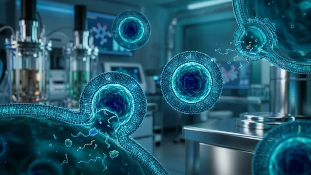
Cells constantly communicate. They send tiny messages to each other. These messages travel inside microscopic bubbles. These bubbles are called extracellular vesicles. Think of them like biological mail trucks. Exosomes are one specific type of these tiny trucks. They are incredibly small. Billions could fit on a pinhead.
All exosomes carry cargo. This cargo includes proteins, lipids, and genetic material like RNA. This cargo can change the behavior of a receiving cell. It can tell a cell to grow, repair, or even calm down. But not all exosomes are the same. Their cargo depends on the parent cell. A stem cell’s exosomes differ from an immune cell’s exosomes.
This is where exoe exosomes enter the story. The term “Exoe” points to a special source. These exosomes come from particular types of cells grown outside the body. These source cells are chosen for their potent natural abilities. Scientists carefully control their environment. This process optimizes the exosomes they release.
So, what makes Exoe exosomes distinct? It is a combination of source and consistency. First, the parent cells are selected for their therapeutic potential. Second, their production is standardized. This means each batch has a similar, defined profile of beneficial cargo. They are not just generic exosomes from any cell.
Compare it to juice. Regular exosomes are like juice squeezed from random fruit. The result is unpredictable. Exoe exosomes are like juice from a specific, nutrient-rich fruit variety. It is grown in optimal soil and harvested at peak ripeness. The final product is consistently powerful and reliable.
Their unique profile gives them exciting advantages. They often carry higher levels of key signaling molecules. These molecules can instruct cells to reduce inflammation. They can promote tissue repair more effectively. They can support the body’s own healing systems with remarkable precision.
Why should you care about this distinction? For science, it means a more reliable tool for research and therapy. For medicine, it promises treatments based on consistent, natural cellular messages. Understanding this difference is crucial. It separates hopeful generalities from targeted, potent science.
The next step is seeing how these vesicles are actually made and collected for use.
The Growing Importance of Exoe Exosomes in Science
Scientists are finding exosomes everywhere. They float in blood, urine, and spinal fluid. These tiny bubbles act as cellular messengers. For years, studying them was difficult. The exosomes from blood samples were a messy mix. They came from many cell types at once. This made results confusing and hard to repeat.
Exoe exosomes solve this critical problem. Their controlled production creates a clean, uniform tool. Researchers can now run precise experiments. They can trace specific effects back to a known vesicle. This reliability accelerates discovery. It turns vague observations into solid data.
Consider disease detection. Sick cells often release altered exosomes. A tumor might send out vesicles that help it spread. In a mixed sample, these dangerous signals get lost. But scientists can design exoe exosomes to mimic disease markers. They use them to calibrate super-sensitive tests. The goal is a simple blood test for early cancer warning.
Therapeutic potential is even more compelling. Medicine is shifting from blunt tools to targeted messages. Drugs often affect the whole body and cause side effects. Exoe exosomes offer a different approach. They carry natural instructions to specific cells.
Think of them as precision-guided medical packages. Their membrane protects the cargo. It also helps them find the right cellular address. Researchers are packing them with beneficial molecules. – They can carry signals to calm an overactive immune system. – They can deliver growth factors to repair heart tissue after an attack. – They can cross the blood-brain barrier, a huge hurdle for most drugs.
This opens doors for treating brain diseases.
The commercial interest is logical. Consistent products enable consistent therapies. Investors and companies see a path from the lab to the clinic. Funding is flowing into this space. It supports larger studies and better manufacturing technology. The field is moving fast from basic science to applied solutions.
However, importance brings responsibility. With powerful tools come big questions. Scientists must prove these vesicles are safe and effective in humans. They must scale up production without losing quality. The growing importance sets the stage for the next big challenge: turning promise into proven practice.
The journey from a research concept to a real treatment is complex. It requires rigorous testing and clever engineering. The next phase involves mastering how these potent vesicles are prepared for real-world use.
Key Differences Between Exoe Exosomes and Regular Exosomes
All cells release tiny bubbles called exosomes. They are messengers in our bodies. But not all exosomes are the same. Exoe exosomes are a special group. They stand out for their purity and power.
Think of regular exosomes as general mail. They carry many types of messages. Their contents can be mixed. Exoe exosomes are more like priority courier packages. They are selected for a specific job. Scientists define them by their origin and contents.
Their source is the first key difference. Regular exosomes come from many cell types during normal activity. Exoe exosomes often come from cells grown under special conditions. These conditions make the cells produce a superior vesicle. The cells are guided to pack them with helpful molecules.
The second difference is consistency. A batch of regular exosomes can be unpredictable. It may contain different amounts of cargo. An Exoe exosome batch is highly uniform. Each vesicle is more alike. This reliability is crucial for medicine. Doctors need every dose to act the same way.
Their membrane structure is also distinct. All exosomes have a protective lipid layer. The Exoe exosome membrane has particular proteins. These proteins act like precise mailing labels. They help the vesicle find its target cell with great accuracy. This improves delivery and reduces side effects.
Finally, their cargo is carefully controlled. Regular exosomes carry a natural mix of signals. – Exoe exosomes can be engineered to carry more of a specific therapeutic molecule. – They might contain a concentrated dose of healing microRNAs. – They could be loaded with enzymes to fix a cellular error.
This intentional design makes them potent tools.
Why should you care about these differences? Uniformity leads to safer treatments. Targeted delivery means stronger effects with fewer problems. The unique traits of Exoe exosomes solve major hurdles in drug development. They turn a natural process into a refined technology.
Understanding this sets the stage for the next step. We know what makes them special. Now we must explore how scientists actually create these precise vesicles in the lab.
How Exoe Exosomes Are Discovered in the Body
Scientists first found clues about Exoe exosomes by studying sick cells. They noticed something strange. Some cells under severe stress released special vesicles. These vesicles were different from common exosomes. They had a unique molecular signature. This signature was like a fingerprint. Researchers could track it in blood and other fluids.
The body creates Exoe exosomes during specific states. These are not everyday events. Think of them as emergency signals or precision tools. Their natural production is a focused response. It happens in three main situations.
- During intense cellular repair after major injury.
- In certain advanced stages of immune system activation.
- As part of highly coordinated developmental processes.
Their discovery required new tools. Regular exosome analysis was not enough. Scientists used advanced sorting technologies. These machines can separate tiny particles based on surface markers. The unique protein label on the Exoe membrane was the key. It allowed researchers to isolate them for the first time. This was a major breakthrough.
So what is their natural job? Their role is about high-stakes communication. Normal exosomes handle daily cellular chatter. Exoe exosomes carry urgent, specific commands. For example, a damaged tissue might release them. These vesicles travel to stem cells in bone marrow. They deliver a precise signal for repair. This starts a powerful healing process.
Their cargo explains their potency. In nature, they don’t carry random mixes. They are packed with selective molecules. A natural Exoe exosome might contain a concentrated set of instructions. It tells a target cell to change its behavior immediately. This could mean stopping inflammation or starting regeneration.
Finding them in the body is like discovering a new postal service. The regular mail delivers letters and bills. This special service delivers critical government orders. The system uses different envelopes and faster routes. The discovery proved that cells already make superior vesicles sometimes. Science is now learning to control that process.
Why does this natural origin matter? It shows these vesicles are not just a lab invention. The body already uses this sophisticated system for important tasks. This gives scientists a blueprint. They mimic and improve upon a natural design. Understanding where Exoe exosomes come from helps us see their true potential. It connects biology’s wisdom to modern medicine’s goals.
This foundation leads to an obvious question. How do researchers move from observing these vesicles to making them? The next step involves sophisticated laboratory techniques and careful design principles.
Why Exoe Exosomes Matter for Future Health Solutions
Exoe exosomes offer a new way to deliver medicine. Think of them as ultra-smart delivery trucks. These trucks know the exact address in the body. They also carry a precise package. This could change how we treat many diseases.
Current drugs often flood the entire system. They go everywhere. This causes side effects. The medicine dose must be high to reach the right spot. Exoe exosomes solve this problem. They deliver their cargo directly to sick cells. This makes treatment more powerful. It also makes it safer.
Their potential spans several major health challenges. One key area is fighting cancer. Tumors are clever at hiding from our immune system. Researchers can load Exoe exosomes with specific signals. These signals can teach immune cells to see and attack the cancer. This approach is called immunotherapy. It is already showing great promise. Exoe exosomes could make it even more targeted.
Another critical field is regenerative medicine. This means healing damaged tissues. Heart muscle after an attack is one example. Brain cells after a stroke are another. These tissues heal poorly on their own. Exoe exosomes can carry instructions to reduce scar tissue. They can also tell local cells to grow new, healthy ones. This is not science fiction. Early animal studies show real repair.
Chronic inflammation is at the root of many illnesses. Arthritis and some bowel diseases are linked to it. Today’s drugs can suppress the whole immune system. This leaves patients open to infections. Exoe exosomes could be designed for a smarter job. They might tell only the overactive immune cells to calm down. This would stop the damage without harmful side effects.
The benefits of this technology are clear and specific. – Precision: They target only diseased cells, sparing healthy ones. – Natural Design: The body recognizes them more easily than synthetic particles. – Potency: A small dose can have a large effect due to efficient delivery. – Versatility: They can carry different types of cargo, from RNA to proteins.
Why should you care about these tiny vesicles? Their impact goes beyond single treatments. They represent a shift in medical thinking. Medicine is moving from broad therapies to precise fixes. Exoe exosomes are tools for this new era. They use the body’s own language to promote healing.
This matters for future health solutions because it promises better outcomes. Treatments could become more effective and less harsh. Recovery times could shorten. Conditions we see as permanent today might become treatable tomorrow. The goal is not just to manage disease, but to reverse its damage.
The road from lab to clinic still has steps. Scientists must ensure these vesicles are perfectly safe. They must prove they work consistently in large human trials. Manufacturing them in pure, large quantities is another hurdle. Yet, the path is clear. The science is building rapidly.
Understanding Exoe exosomes helps us see this future. It connects a biological discovery to real-world hope for patients everywhere. The next logical question involves the practical journey from a promising idea to an available therapy. This process relies on rigorous testing and scalable production methods that maintain the vesicles’ unique power.
The potential is vast, but realizing it requires careful work and proof.
The Unique Features of Exoe Exosomes
Molecular Signatures That Define Exoe Exosomes
Exoe exosomes carry a special molecular ID card. This ID is not a single molecule. It is a specific combination of many molecules on their surface. Think of it like a passport with many unique stamps. This combination defines them as Exoe exosomes.
All exosomes have some common markers. These are proteins like CD63 and CD81. They confirm a vesicle is an exosome. But Exoe exosomes have extra signatures. These signatures give them unique abilities.
One key feature is their lipid membrane. This outer layer is not a simple bubble. It is rich in certain fats called lipids. These lipids help the exosome fuse with a target cell. This fusion is like a secure handshake. It allows the cargo to be delivered directly inside.
The most important signatures are the proteins. Some proteins sit on the surface. They act as address labels. They guide the exosome to the right cell type in the body. For instance, one protein might target liver cells. Another could seek out skin cells.
Other proteins are inside the vesicle. These are the tools and instructions. They include growth factors and enzymes. These molecules can kick-start repair processes in a recipient cell.
The cargo itself is a major part of the signature. Exoe exosomes often carry specific types of genetic material. This includes microRNAs. These are short strands of RNA that do not make proteins. Instead, they act as control switches. They can turn genes in the target cell on or off.
The exact mix of these components varies. It depends on the parent cell that made them. A stem cell will produce Exoe exosomes with a different signature than an immune cell. The parent cell’s state also matters. A healthy cell and a stressed cell release different vesicles.
Scientists identify these signatures using advanced tools. One common method is flow cytometry. This machine can detect surface proteins on tiny particles. Another technique is mass spectrometry. It can list all the proteins inside a sample of exosomes.
Why do these molecular details matter? They explain how Exoe exosomes work. The surface proteins ensure delivery to the correct location. The internal cargo then executes a precise program. This two-step process is efficient and natural.
These signatures also serve as a quality check. Researchers can test a batch of exosomes. They look for the expected molecules. Finding them confirms the vesicles are genuine and potent. Missing signatures mean the batch might not work as intended.
Finally, these molecules offer a path for engineering. Scientists can learn from nature’s designs. They might add extra targeting proteins to improve delivery. They could also load specific therapeutic RNAs inside the vesicles.
Understanding these molecular blueprints is crucial. It moves us from seeing Exoe exosomes as simple bubbles to recognizing them as sophisticated messengers. Their power lies in this precise chemical makeup, which dictates their journey and their effect in the body.
This leads to the next practical challenge: how to produce these complex vesicles consistently at a large scale while preserving their delicate molecular signatures, a task essential for turning their potential into real treatments.
Biophysical Properties of Exoe Exosomes
Exoe exosomes are incredibly small. Their size is measured in nanometers. One nanometer is one billionth of a meter. For scale, a single sheet of paper is about 100,000 nanometers thick. Most Exoe exosomes fall into a tight range. They are typically between 60 and 80 nanometers wide.
This specific size is not random. It is a direct result of their unique molecular blueprint. The proteins that form their structure control this precise measurement. This compact size gives them key advantages. They are small enough to navigate through tissues. They can also slip into the spaces between cells with ease.
Their shape is another critical feature. Under powerful microscopes, Exoe exosomes appear as perfect spheres. They look like tiny, round bubbles. This spherical shape is essential for their stability in fluids. A smooth, round object moves with less friction. It also resists damage better than an irregular shape would.
The membrane surrounding them is remarkably strong. Think of it like a durable plastic bag. It is a lipid bilayer filled with cholesterol and special proteins. This structure protects the precious cargo inside. The cargo includes RNA and signaling proteins. The membrane keeps this material safe from enzymes in the blood that would destroy it.
This toughness allows Exoe exosomes to travel far. They can move through the bloodstream without bursting. They survive harsh conditions that would break other vesicles. Their journey can start in one tissue and end in another distant organ. The membrane ensures the delivery of an intact message.
Their physical properties also affect how they are studied and used. Scientists can separate them from other particles based on size and density. This process is called ultracentrifugation. Machines spin samples at very high speeds. Heavier or larger particles sink first. The Exoe exosomes form a distinct layer that can be collected.
- Size: Enables navigation through tiny biological spaces.
- Shape: Promotes stability and efficient movement in fluids.
- Membrane Strength: Protects cargo for long-distance delivery.
These biophysical traits work together with their chemical signatures. The right molecules build the right structure. The right structure enables the right function. You cannot have one without the other. A perfect molecular code would be useless inside a fragile vesicle that pops immediately.
Understanding these physical traits solves practical problems. It tells engineers how to handle these vesicles in a lab. It shows why they can be stored under certain conditions. It explains their potential as reliable carriers for medicines. Their natural durability is a major part of their promise.
In essence, Exoe exosomes are nature’s own precision-designed nanoparticles. They have an ideal size for delivery, a stable shape for travel, and a resilient membrane for protection. These are not fragile bubbles. They are robust, sophisticated couriers built for a critical mission within the body’s vast landscape.
How Exoe Exosomes Carry Information Between Cells
Exoe exosomes act as tiny cellular messengers. They carry precise instructions from one cell to another. This process is fundamental to health and disease. A single vesicle can change a recipient cell’s behavior.
The cargo inside determines the message. Exoe exosomes pack different types of molecular information. This cargo is carefully selected by the parent cell. Think of it as loading a secure USB drive with specific files.
The main types of cargo include: – Proteins: These can be enzymes that start chemical reactions. They can also be signals that latch onto a cell’s surface. – Lipids: These molecules are part of the exosome’s own membrane. They can fuse with a target cell. – Nucleic Acids: This is the most direct genetic instruction. RNA is a common payload.
MicroRNAs are a key example. These are short strands of genetic material. They do not carry blueprints for proteins. Instead, they control which genes in the target cell are active or silent. An Exoe exosome from a stem cell might contain microRNAs that tell a skin cell to repair itself. A cancer cell’s exosome might carry microRNAs that shut down immune defenses.
Delivery is a precise operation. The journey starts with release from the parent cell. The exosome travels through bodily fluids. It navigates to a specific destination. The surface of the Exoe exosome has address labels. These are proteins and sugars that match receptors on certain target cells.
Binding is the first step. The exosome docks onto the target cell’s membrane. Then, one of two primary delivery methods occurs. The vesicle can fuse directly with the cell’s outer layer. This dumps the cargo straight into the cell’s interior. Alternatively, the whole exosome can be swallowed by the cell. The cell membrane wraps around and engulfs the vesicle.
Once inside, the cargo gets to work. Proteins may start new processes. Lipids can become part of the cell’s own structure. Delivered RNA molecules head to the cell’s control center. They then influence which proteins the cell makes. This changes the cell’s function without altering its core DNA.
This system is incredibly efficient. One sending cell can dispatch many exosomes. Each Exoe exosome can influence multiple target cells. The signal amplifies quickly across tissues. It allows for coordinated body-wide communication.
The information flow is two-way. Cells constantly send and receive these packets. A healthy state depends on balanced signaling. Disease often involves corrupted messages. For instance, tumors use exosomes to prepare distant organs for cancer spread. They send signals that create a welcoming environment.
Understanding this cargo system is crucial for medicine. If we know the message, we can block a harmful one. We can also mimic a helpful one. Scientists could load therapeutic Exoe exosomes with specific RNAs or drugs. These engineered couriers would use the body’s own natural delivery network.
In summary, Exoe exosomes are more than just carriers. They are active communication devices. They transfer functional biological software between cells. This elegant system ensures tissues work in harmony. The next question is how this natural process can be guided for healing.
The Role of Exoe Exosomes in Cellular Communication
Exoe exosomes act like targeted text messages between cells. They are not random broadcasts. A skin cell sends different exosomes than a nerve cell. Each type carries a unique molecular address. This address ensures the vesicle finds the correct recipient cell. Think of it as a cellular zip code.
The surface of an Exoe exosome is covered with proteins. These proteins act as both an address label and a key. They bind to specific receptors on a target cell’s membrane. This binding is the first step in communication. It is a precise lock-and-key mechanism. Only the right key opens the door for the next step.
This targeting prevents chaos. Without it, signals would go everywhere. A liver signal might accidentally trigger a brain cell. Exoe exosomes maintain order in this complex system. Their directed delivery makes communication efficient and meaningful.
Once docked, the exosome delivers its instructions. The cargo inside tells the recipient cell what to do. The commands are clear and direct. Common instructions include: – Grow and divide more. – Move to a new location. – Change your primary function. – Activate your defense systems. – Prepare for repair work.
The recipient cell does not question these orders. It follows them precisely. This allows tissues to respond quickly to changes. For example, a muscle tear releases signals. Nearby stem cells receive Exoe exosome messages. They then travel to the injury site and begin repairs.
The speed of this system is remarkable. Signals travel quickly through bodily fluids. An exosome released in one organ can reach another in minutes. This creates a rapid-response network across the entire body. It is faster than waiting for hormonal signals in the blood.
Exoe exosomes also carry historical data. Their cargo reflects the health of their parent cell. A stressed cell sends different exosomes than a healthy one. The recipient cell reads this context. It adjusts its response based on the sender’s condition. This adds a layer of sophistication to the conversation.
Cancer cells exploit this natural system. They send deceptive Exoe exosomes with corrupted addresses and false commands. These malicious messages can tell blood vessels to grow toward a tumor. They can also tell immune cells to stand down. This hijacking shows the power of precise cellular communication.
Researchers aim to intercept bad messages. They also want to send new therapeutic ones. Scientists could engineer Exoe exosomes with custom addresses and cargo. These designed vesicles could deliver repair commands directly to damaged heart tissue after an attack. They could tell overactive immune cells in arthritis patients to calm down.
The true uniqueness of Exoe exosomes lies in this combination of precision and power. They deliver potent biological instructions with pinpoint accuracy. This makes them ideal for natural healing and advanced medicine. Their role transforms cellular chatter into a coordinated dialogue that maintains our health every single day. Understanding this dialogue is the first step toward mastering it for therapy.
Why Exoe Exosomes Are More Stable Than Other Vesicles
Exoe exosomes possess a remarkable trait. They last much longer in the bloodstream than many other biological carriers. This stability is not an accident. It results from specific engineering at the cellular level. Think of a regular soap bubble. It pops quickly in air. Now imagine a tiny, robust capsule with a reinforced shell. That capsule is the Exoe exosome. Its longevity allows it to travel far from its origin cell. It can deliver its message to a distant target without falling apart.
Their membrane structure is key to this durability. All exosomes have a lipid bilayer. Yet Exoe exosomes have a unique composition. Their membrane is enriched with special lipids and proteins. These components are tightly packed together. This creates a sturdy barrier. It protects the delicate cargo inside from the harsh environment outside. The body’s bloodstream is full of enzymes that can break things down. It also has forces that can tear fragile vesicles apart. The Exoe exosome’s tough shell resists these attacks.
Surface molecules also play a major role. Exoe exosomes carry specific proteins on their outside. These proteins do more than just act as an address label. Some of them interact with proteins in the blood plasma. This interaction forms a protective coating around the vesicle. It is like a shield. This shield makes the exosome less visible to the body’s cleanup systems. Immune cells are less likely to grab and destroy it. So the vesicle enjoys a longer journey.
Size and shape contribute to stability as well. Exoe exosomes have a very consistent, small size. They are typically between 30 and 150 nanometers in diameter. This is incredibly tiny. Their uniform shape reduces friction as they move through fluids. A rough, irregular particle gets caught more easily. A smooth, round one flows better. This efficient design helps them navigate the circulatory system for extended periods.
Their internal cargo even helps them survive. The molecules inside an Exoe exosome are not just passive passengers. Some nucleic acids and proteins help maintain the vesicle’s structure. They can stabilize the interior environment. This prevents the exosome from collapsing from within. It is a self-sustaining system designed for a long trip.
Compare this to other extracellular vesicles. Some microvesicles are simply buds pinched off from a cell’s membrane. They are often larger and less organized. Their structure is looser. They tend to break down faster. Free-floating proteins or lipids in blood are cleared in minutes or hours. A well-made Exoe exosome can circulate for much longer—sometimes days. This extended window is crucial for biological communication.
Why does this stability matter for medicine? A therapeutic message needs time to find its target. A fragile carrier might degrade before reaching the right cell. An Exoe exosome’s endurance increases its chance of success. Engineers could load them with drugs or healing instructions. These stable capsules would protect their precious cargo all the way to the destination. This makes them reliable messengers for future treatments.
Scientists measure this stability in experiments. They collect Exoe exosomes and place them in conditions that mimic body fluids. They then track how many remain intact over time. The results consistently show their resilience. This proven durability supports their potential for real-world applications where timing is everything.
In summary, Exoe exosomes are built to last. Their robust membrane, protective shield, and optimal design grant them unusual staying power in the body. This fundamental feature underpins their role as precise long-distance messengers. It transforms them from simple bubbles into enduring couriers capable of sophisticated tasks. Their stability ensures the cellular dialogue continues until the message is fully delivered
Advanced Technologies for Studying Exoe Exosomes
Modern Methods to Isolate Exoe Exosomes
Isolating Exoe exosomes is a critical first step for any research. Scientists must separate them from a crowded biological soup. This soup contains cells, debris, and many other particles. The goal is to get a clean sample. A pure sample allows for accurate study of the exosomes’ cargo and effects.
Several modern methods achieve this separation. Each technique uses a different physical property of the vesicles. The choice depends on the sample type and the needed purity. No single method is perfect. Researchers often combine techniques for the best results.
Ultracentrifugation is a traditional and common approach. It uses extremely high spinning speeds. A machine called an ultracentrifuge spins the sample. This creates powerful forces. Heavier particles like cells get forced to the bottom first. Lighter particles like Exoe exosomes settle out later. The process is time-consuming. It requires expensive equipment. However, it can process large sample volumes.
Size-based techniques are a popular alternative. These methods filter the sample. One common tool is size-exclusion chromatography. The sample flows through a column filled with porous beads. Smaller particles get trapped in the pores. They take longer to exit. Larger particles flow around the beads quickly. Exoe exosomes, having a specific size range, elute in a predictable fraction. This method is gentle. It helps preserve the exosome structure.
Another approach uses immunoaffinity capture. This method targets surface markers. Exoe exosomes have unique proteins on their outer membrane. Scientists use antibodies that stick to these proteins. The antibodies are attached to magnetic beads or a surface. When the sample passes by, the Exoe exosomes bind. Everything else gets washed away. This gives very pure samples. It is excellent for studying specific exosome types. But it can be costly and may miss exosomes without the target marker.
Precipitation kits offer a simpler protocol. Chemicals are added to the sample. These chemicals change the solution’s conditions. They reduce the solubility of vesicles. The Exoe exosomes clump together and fall out of solution. The pellet is then collected. This method is fast and requires no special machinery. It works with many starting materials. The downside can be lower purity. Other molecules may precipitate alongside the vesicles.
Microfluidic devices represent cutting-edge technology. These are tiny chips with etched channels. The sample flows through these microscopic pathways. Engineers design traps or filters within the channels. These features catch particles of a certain size or charge. Exoe exosomes get directed to one outlet. Other components go to waste channels. This method is fast and uses very small sample volumes. It shows great promise for clinical diagnostics.
Choosing the right isolation strategy is foundational. The method impacts everything that follows. It affects the quality and reliability of all downstream data. Researchers carefully balance speed, cost, yield, and purity needs.
Successful isolation unlocks the next phase of analysis. Scientists can then probe the contents and functions of these robust messengers. This clean separation is what allows their detailed study and future application.
The field continues to evolve towards faster, integrated systems. The ideal future method would be quick, affordable, and highly precise. For now, understanding these core techniques reveals how science captures these elusive cellular couriers for closer examination
Analytical Techniques for Exoe Exosome Characterization
Once scientists have a clean sample of Exoe exosomes, the real detective work begins. They need to know exactly what they have captured. Characterization answers critical questions. How big are these vesicles? What is on their surface? What cargo do they carry inside?
A first basic step is sizing and counting. Nanoparticle Tracking Analysis, or NTA, is a common tool. A laser beam shines through the liquid sample. The tiny Exoe exosomes scatter the light. A camera records these flashes of light. Software then tracks the movement of each flash. This movement reveals the particle’s size. The method also provides a count of particles per milliliter. It confirms the isolation was successful.
To see the actual shape and structure, scientists use electron microscopy. A Transmission Electron Microscope, or TEM, is powerful. It uses a beam of electrons instead of light. The sample is prepared on a tiny grid. It is stained with heavy metals for contrast. The electron beam passes through it. This creates a highly detailed black-and-white image. Scientists can see the classic cup-shaped morphology of dried exosomes. They can verify the vesicles are intact and not broken.
Surface markers are like identification badges. Flow cytometry can analyze them. In this method, the sample is stained with fluorescent antibodies. Each antibody sticks to one specific protein on the vesicle surface. The fluid stream passes single particles by a laser. Detectors measure the fluorescent light emitted. This reveals which proteins are present. It helps confirm the vesicles truly are Exoe exosomes with their unique surface signature.
Understanding the molecular cargo is crucial. Scientists often crack the vesicles open to analyze their contents.
- For proteins, they use Western Blotting. Proteins are separated by size on a gel. They are transferred to a membrane. Specific antibodies then bind to target proteins. A reaction produces a visible band. This confirms the presence of key cargo proteins.
- For RNA, techniques like qPCR are used. The genetic material is extracted and copied millions of times. This allows scientists to measure specific RNA sequences. They can see what messages the exosome carries.
- For lipids, mass spectrometry is a key tool. It measures the mass of different molecules. It can identify the unique lipid makeup of the vesicle membrane.
A powerful modern approach is multi-omics. This means analyzing all data types together. Scientists combine protein, RNA, and lipid profiles. They use advanced bioinformatics software. This creates a complete functional portrait of the exosome population. It shows how the pieces work together as a system.
Each technique has trade-offs. Some need large sample amounts. Others are very expensive or slow. Some only give bulk data for millions of vesicles at once. Newer single-vesicle analysis methods are emerging. These will show differences between individual Exoe exosomes in a sample.
This detailed characterization is essential. It moves from simply having particles to truly understanding them. It validates their identity and hints at their potential function. This foundational knowledge directly enables the next step: testing how these robust vesicles actually behave and influence other cells.
How Imaging Reveals Exoe Exosome Structures
Seeing an exoe exosome is not easy. These particles are far smaller than a human cell. They are measured in nanometers. One nanometer is one billionth of a meter. Advanced microscopes are needed to view them. These tools do more than take pictures. They reveal the shape, size, and surface details of individual vesicles. This visual data confirms their physical identity.
Transmission electron microscopy is a classic method. It uses a beam of electrons instead of light. Samples must be prepared carefully. They are stained with heavy metals like uranium. This stain highlights different structures. The electron beam passes through the thin sample. It creates a detailed black-and-white image. The result shows the classic cup-shaped structure of many exosomes. Scientists can see the lipid bilayer membrane clearly. They can also measure the diameter of each vesicle precisely.
Cryo-electron microscopy is a newer, gentler technique. Samples are frozen extremely fast. This traps them in a layer of clear ice. The freezing happens so quickly that water doesn’t form crystals. The structure is preserved in a near-native state. No harsh stains are needed. The vesicles are viewed while still frozen. This method shows the exosomes in their natural rounded shape. It provides incredible detail of surface proteins and complexes.
Atomic force microscopy is a different approach. It does not use a beam. It uses a tiny mechanical probe. This probe scans across the sample surface. It feels the shape of each particle. It moves up and down over bumps and valleys. This movement is tracked by a laser. A computer then builds a three-dimensional map. This map shows the height and width of each exoe exosome. Scientists get precise topographic data. They can see surface roughness and texture.
Super-resolution fluorescence microscopy breaks a old rule. Traditional light microscopes cannot see objects this small. Their resolution limit is about 200 nanometers. Many exosomes are smaller than that. Super-resolution techniques use clever tricks with light. They make fluorescent molecules blink on and off. A computer records thousands of images. It then reconstructs a final picture with much finer detail. This allows scientists to see specific proteins on the exosome surface. They can watch how these vesicles interact with cells in real time.
Each imaging method has strengths. – Electron microscopy gives the highest resolution detail. – Cryo-EM preserves natural shapes without artifacts. – Atomic force microscopy provides 3D height data. – Super-resolution shows specific proteins in living contexts.
Often, scientists combine several techniques. They use one for overall shape and another for protein location. This multi-modal approach builds a complete visual model. It answers critical questions about structure. Is the vesicle intact? What is its exact size distribution? How do proteins arrange on its surface? This structural knowledge complements biochemical data perfectly. Together, they form a robust profile of these promising vesicles, setting the stage for functional experiments that test their capabilities directly in biological systems.
The Use of Sequencing in Exoe Exosome Research
Sequencing reads the biological messages inside exoe exosomes. These vesicles carry more than just proteins. They contain genetic material. This cargo includes RNA molecules. Cells pack these molecules into vesicles for delivery. Scientists can extract and read this RNA. This reveals what the original cell was doing. It shows what signals it was sending.
The process starts with isolation. Researchers first collect a pure sample of exoe exosomes. They use methods like ultracentrifugation or filtration. Next, they break open the vesicle membranes. This releases the RNA inside. The RNA is then converted into DNA. This step uses an enzyme called reverse transcriptase. The resulting DNA is compatible with sequencing machines.
Modern sequencing platforms are powerful. They can read millions of RNA fragments at once. This is called high-throughput sequencing. It provides a massive snapshot of genetic content. The data answers key questions. What types of RNA are present? How much of each type exists? Are there unique genetic signatures?
Two main RNA types are often found. – MicroRNAs are small regulators. They can silence genes in target cells. – Messenger RNAs carry blueprints for making proteins.
Finding these molecules is significant. It means exoe exosomes can change cell behavior directly. They can deliver new instructions to recipient cells.
Data analysis is a major part of the work. Raw sequencing output is a list of genetic codes. Bioinformaticians use software tools. They map each sequence to a known genome. They count how many times each sequence appears. This quantifies expression levels. Sophisticated comparisons can then be made.
Researchers compare exosomes from different sources. They might contrast healthy and diseased cells. They look for RNAs that are only present in one group. These unique molecules become potential biomarkers.
For example, certain microRNAs are abundant in tumor-derived exosomes. Their presence in a blood sample could signal early cancer. This non-invasive detection is a major research goal. Sequencing makes this discovery possible.
The technique also studies exoe exosome function. Scientists can track delivered RNA. They sequence the contents of a recipient cell after exosome exposure. They check if new RNA sequences appear. This confirms successful transfer. It proves the vesicles act as genetic messengers.
Challenges remain in this field. RNA amounts inside a single exosome are tiny. Technical artifacts can contaminate samples. Careful controls are essential for valid results.
Despite hurdles, sequencing is transformative. It moves research from “what they look like” to “what they do.” Genetic analysis uncovers the active communication code within these vesicles. This molecular understanding directly enables the next phase of research: engineering these natural carriers for precise therapeutic goals.
Emerging Tools for High-Throughput Exoe Exosome Analysis
Traditional exosome analysis can be slow. Scientists often study them one sample at a time. New high-throughput methods change this. They let researchers process hundreds of samples in a single experiment. This speed is crucial for discovery.
High-throughput analysis relies on automation and miniaturization. Robots handle liquid transfers. Tiny reaction chambers replace large tubes. This allows many tests to run in parallel. It saves time and precious samples.
One key technology is multiplexed flow cytometry. Special machines analyze exosomes as they flow in a stream. Each vesicle passes a laser beam. The machine can count them and measure surface markers. Advanced systems now tag exosomes with different fluorescent colors. This lets scientists track dozens of targets at once from a single sample.
Another tool is array-based platforms. These are chips with thousands of microscopic spots. Each spot holds a capture antibody for a specific exosome protein. A sample flows over the chip. Exoe exosomes stick to their matching spots. A scanner then reads the entire array quickly. It generates a massive protein profile.
These methods excel at finding patterns. Researchers can compare exosomes from many patients rapidly. They search for common signatures linked to a disease. This scale was impossible with older, slower techniques.
Digital PCR is another high-throughput advance. It partitions a sample into thousands of nanodroplets. Each droplet acts as a separate reaction chamber. This allows absolute counting of RNA molecules from exosomes. It is extremely sensitive and quantitative.
The data from these tools is vast. Bioinformatics becomes essential again. Powerful software spots trends humans would miss. It connects complex molecular data to clinical outcomes.
The benefits of high-throughput analysis are clear: – It accelerates biomarker discovery. – It enables large-scale clinical validation studies. – It provides statistical power for rare exosome subpopulations. – It reduces costs per data point significantly.
For exoe exosomes, this speed is transformative. Their unique biology can now be surveyed across huge populations. Researchers can ask new questions. They can study how exoe exosome signals change over time during treatment. They can screen thousands of compounds to find ones that alter exosome release.
These technologies also face challenges. The equipment is expensive and complex. Generating so much data requires robust storage and analysis plans. Standardizing protocols across labs is an ongoing effort.
Yet the direction is set. The field is moving toward faster, richer, and more comprehensive analysis. High-throughput tools turn exoe exosomes from objects of detailed study into sources of big data. This flood of information is the next step toward reliable diagnostics and personalized therapies based on these vesicles. It builds directly on the foundational science explored earlier, scaling discovery to meet medical needs.
Exoe Exosomes in Diagnostics and Therapy
How Exoe Exosomes Improve Disease Detection
Exoe exosomes carry molecular messages from their parent cells. If a cell becomes sick, its exoe exosomes change. This offers a powerful way to spot disease long before symptoms appear.
Think of them as tiny diagnostic reporters. They travel through blood and other easy-to-access fluids. Doctors call this a “liquid biopsy.” It is far simpler than a tissue biopsy. A standard blood draw can provide these vesicles.
Their unique surface proteins are the first clue. Exoe exosomes from cancer cells often have different surface markers. These markers act like flags. A test can scan a blood sample for these specific flags. Finding them suggests a tumor is present somewhere.
The cargo inside is even more telling. Exoe exosomes from diseased cells contain abnormal molecules. This includes specific microRNAs and proteins linked to illness. For example, pancreatic cancer exoe exosomes may carry a distinct RNA signature. Detecting this signature can signal early-stage cancer.
This early detection is crucial for diseases like Alzheimer’s. Brain cells release exoe exosomes into cerebrospinal fluid. These vesicles can contain tau proteins or beta-amyloid fragments. Finding these molecules gives a window into brain health decades before memory loss.
The process for using them in detection has clear steps. – First, a sample is taken from a patient. This is usually blood or urine. – Next, exoe exosomes are separated from the fluid. – Then, scientists analyze their surface markers and internal cargo. – Finally, they compare the findings to known disease profiles.
Exoe exosomes improve detection in several key ways. They are abundant and stable in circulation. They protect their molecular cargo from degradation. They come directly from the tissue of interest, like a brain or pancreas. This provides precise information about that organ’s state.
Sensitivity is a major advantage. These vesicles can reveal very small changes in cell biology. A tiny group of precancerous cells can release enough exoe exosomes to be found. This allows for intervention at the most treatable stage.
Specificity is another benefit. The signature of an exoe exosome from a lung tumor differs from one linked to liver disease. Tests can be designed to look for very precise signals. This reduces false alarms and pinpoints the problem.
Monitoring treatment becomes possible too. After therapy begins, the type and number of exoe exosomes should shift. A decrease in cancer-associated vesicles suggests the treatment is working. Doctors can track this change over time with simple blood tests.
Challenges remain, of course. Scientists must ensure tests are reliable and consistent. They must confirm which signals are truly linked to early disease. Large clinical trials are needed to validate these approaches.
Yet the potential is immense. Exoe exosomes turn routine blood work into a detailed health report. They move medicine from reacting to symptoms to preventing full disease onset. This proactive approach could save countless lives.
The natural next question is therapeutic use. If we can find diseases with these vesicles, can we also treat them with them?
Exoe Exosomes as Biomarkers for Various Conditions
Exoe exosomes act as precise messengers from troubled tissues. Their molecular cargo changes when a cell becomes diseased. This provides a clear signal in a patient’s blood.
Cancer detection is a primary focus. Tumors release large numbers of these vesicles. Their cargo includes proteins and RNA fragments unique to cancer. For instance, exoe exosomes from pancreatic tissue may carry a protein called GPC1. Finding this marker suggests a pancreatic tumor might be present. This can happen long before a scan shows any mass.
Neurological disorders also leave traces. The brain is protected by a tight barrier. Large molecules cannot cross it easily. However, exoe exosomes from neurons can pass into the bloodstream. They may carry signs of Alzheimer’s disease, like tau protein. Analyzing these vesicles offers a window into brain health without invasive procedures.
Inflammatory and autoimmune diseases show distinct patterns too. Conditions like rheumatoid arthritis cause joint inflammation. Immune cells in the joints release exoe exosomes filled with inflammatory signals. These signals can be measured. Doctors can then gauge how severe the inflammation is internally.
The process for using these biomarkers involves several steps. – First, a blood sample is taken from a patient. – Next, all exosomes are separated from the plasma. – Then, tools like antibodies capture only the exoe exosomes from a specific organ. – Finally, scientists analyze the captured vesicles for disease markers.
This approach is far more targeted than traditional tests. Standard blood work measures general substances like liver enzymes. High enzymes indicate liver stress but not the exact cause. Exoe exosomes from the liver would provide more detail. Their cargo could point directly to fatty liver disease, viral hepatitis, or early cancer.
Cardiovascular health is another promising area. Stressed heart muscle cells release specific exoe exosomes. These vesicles contain molecules linked to cardiac strain. Monitoring them could warn of a potential heart attack weeks in advance. It allows for preventive treatment to be started.
The true power lies in combination. A single blood draw could be screened for exoe exosomes from multiple organs simultaneously. A profile might show normal brain markers, elevated inflammatory joint signals, and subtle pancreatic warnings. This creates a holistic health snapshot.
Researchers are validating these signals across many conditions. They study large groups of people over time. The goal is to confirm which cargo changes are reliable early warnings. This work is complex but critical for future clinical use.
Exoe exosomes transform vague symptoms into precise biological data. A patient’s fatigue could be linked to an immune signal from their exoe vesicles. Unexplained weight loss might correlate with specific metabolic RNA found in these particles. Medicine moves closer to objective, early detection.
This foundational role in diagnosis naturally leads to a therapeutic question. If we can intercept these disease signals, can we also engineer the vesicles to carry treatments back?
Therapeutic Applications of Exoe Exosomes
The same exoe exosomes that signal disease can be engineered to treat it. Scientists are turning these natural carriers into precision delivery vehicles. They can load therapeutic cargo directly inside the vesicles. This cargo then travels straight to the organ or cell type it came from.
Think of it as a targeted return trip. A liver-derived exoe exosome naturally seeks other liver cells. Researchers can fill it with healing molecules. These could be small drugs, protective proteins, or corrective RNA. The exosome delivers its payload directly to the diseased liver tissue. This method spares healthy organs from side effects.
The process involves several key steps. First, scientists collect exoe exosomes from specific cells grown in the lab. Next, they load the vesicles with the chosen treatment. This is done using gentle electrical pulses or incubation. Finally, the engineered exosomes are purified and prepared for use.
Therapeutic exoe exosomes show great promise for tough conditions. In neurodegenerative diseases like Alzheimer’s, they could carry growth factors across the blood-brain barrier. For damaged heart tissue after an attack, they might deliver instructions to reduce scarring. In autoimmune disorders, they could be designed to calm an overactive immune response precisely where it attacks.
Cancer therapy is a major focus. Tumors release many exoe exosomes. These vesicles naturally home back to cancer cells. Engineers can load them with chemotherapy drugs or molecules that trigger cell death. This turns the cancer’s own communication system against it. It allows for lower drug doses with stronger effects on the tumor.
Regenerative medicine also benefits from this approach. Exoe exosomes from stem cells are packed with healing signals. They can instruct damaged tissues to repair themselves. For example, exosomes from mesenchymal stem cells can reduce inflammation in arthritic joints. They can also promote the growth of new blood vessels in injured muscles.
The advantages over conventional treatments are clear. – Targeted Delivery: Medicines go straight to sick cells. – Reduced Toxicity: Healthy tissues experience fewer side effects. – Natural Carrier: The body’s own vesicles are less likely to trigger an immune reaction. – Multi-Cargo Capacity: One exosome can carry different types of healing molecules at once.
Current research is solving practical challenges. Scientists are working on large-scale production methods. They are improving techniques to load cargo efficiently. Studies also focus on extending the circulation time of therapeutic exosomes in the bloodstream. Each solved problem brings this technology closer to clinics.
Safety remains a top priority. Researchers carefully check that engineered exoe exosomes do not cause unintended effects. They monitor for any potential immune system activation. Long-term studies in animals help establish safe profiles before human trials begin.
The future vision is personalized medicine. A patient’s own cells could provide the source for their therapeutic exoe exosomes. These custom vesicles would be loaded with drugs tailored to their specific disease profile. This approach maximizes treatment effectiveness and safety.
The journey from diagnostic signal to therapeutic agent is now underway. Exoe exosomes offer a two-way street for medicine—first revealing disease, then treating it with unmatched precision. This dual capability positions them as a cornerstone for the next generation of medical care, moving us beyond simple symptom management to true cellular repair.
Exoe Exosomes in Targeted Drug Delivery Systems
Exoe exosomes act as nature’s perfect delivery trucks. Their natural design solves a major drug delivery problem. Medicines often struggle to reach the right cells. They can break down in the bloodstream. They might also harm healthy tissues. Exoe exosomes provide a protective container. They shield their cargo until arrival.
Scientists load these vesicles with therapeutic cargo in several ways. One method is called incubation. Purified exoe exosomes are mixed with drug molecules. The cargo slowly passes through the vesicle membrane. Electroporation is another common technique. A brief electric pulse creates tiny pores in the exosome’s surface. Drugs quickly enter through these temporary holes. After the pulse, the membrane seals itself. The cargo is trapped safely inside.
The real magic lies in targeting. An exoe exosome needs an address label. Cells display specific proteins on their outer surface. These are like unique door handles. Researchers can engineer exoe exosomes to carry matching proteins. These are called targeting ligands. A common example is a peptide. This peptide seeks out and binds only to its matching handle.
Think of it like a key for a lock. The engineered exoe exosome circulates in the body. It ignores cells without the correct lock. When it finds a cell with the matching protein, it docks. The vesicle then merges with the cell’s membrane. It delivers its healing cargo directly into the cell’s interior. This is targeted drug delivery.
The advantages of this system are clear. – Precision: Drugs go primarily to diseased cells. – Protection: The lipid bilayer guards fragile molecules like RNA. – Efficiency: More medicine reaches the intended target. – Natural Entry: Cells readily accept these biological vesicles.
Cancer therapy is a prime example. Tumor cells often overexpress certain proteins. Scientists can design exoe exosomes to target those markers. They load the vesicles with anti-cancer drugs or toxic RNA molecules. The exoe exosomes then seek the tumor. They deliver their deadly payload directly to cancer cells. This spares surrounding healthy tissue from damage.
Neurological diseases also benefit. The blood-brain barrier blocks most drugs from entering the brain. Exoe exosomes have a natural ability to cross this barrier. They can be loaded with medicine for Alzheimer’s or Parkinson’s disease. Their natural composition helps them slip past biological defenses. They deliver treatment where it is needed most.
Production scalability is an active research area. Growing enough parent cells is the first step. These cells must release vast quantities of exoe exosomes. Scientists use bioreactors for this task. These are large tanks that provide ideal growth conditions. The next step is harvesting and purifying the vesicles. This must be done without damaging their delicate structure.
Future systems may be even smarter. Researchers are designing exoe exosomes that release cargo on command. An external trigger could be used. Light or ultrasound are possible triggers. This would allow doctors to control the exact moment of drug release. Another idea involves logic-gated vesicles. These exoe exosomes would only unload their cargo if two conditions are met at the target cell.
The path from lab to patient involves rigorous testing. Each engineered batch must be characterized. Scientists check its size, charge, and surface markers. They confirm the drug is properly loaded inside. Animal studies then show how it behaves in a living system. These steps ensure safety and effectiveness.
Targeted delivery turns a potent drug into a precise tool. Exoe exosomes provide the guidance system and protective capsule. This merges biological intelligence with therapeutic power, creating a new standard for treatment accuracy that minimizes collateral damage to the patient’s body
Case Studies of Exoe Exosome Use in Medicine
Cancer detection offers a powerful example. Tumors shed exosomes into the bloodstream. These vesicles carry molecular fingerprints from their parent cells. Researchers can analyze these exoe exosomes to find cancer early. A specific surface protein might signal a brain tumor. A unique RNA fragment could indicate pancreatic cancer. This method is like finding a distinct message in a bottle floating in the blood. It is less invasive than a tissue biopsy. It can also monitor how a tumor responds to therapy over time.
Neurological diseases are another key area. The brain is protected by a tight barrier. This barrier blocks many drugs and diagnostic tools. Exosomes from brain cells can cross this barrier naturally. Scientists study exosomes from spinal fluid or blood. They look for signs of Alzheimer’s disease. Clumps of toxic proteins can be carried inside these vesicles. Finding these clumps provides an early warning sign. It helps doctors diagnose the disease before severe symptoms appear.
Therapeutic applications are now in clinical trials. One approach treats inflammatory conditions. Mesenchymal stem cells release exosomes with healing signals. Engineers harvest and purify these exoe exosomes. They then inject them into injured joints. The exosomes reduce swelling and pain. They encourage tissue repair. This use avoids the risks of whole cell therapy.
Another case targets liver fibrosis. This is scarring caused by chronic damage. Researchers load exosomes with special RNA molecules. This RNA silences a gene that drives scar tissue growth. The exosomes deliver their cargo directly to liver cells. This precision stops the scarring process. It allows healthy tissue to regenerate.
Cardiac repair after a heart attack is also being explored. Damaged heart muscle cannot regenerate well. Exosomes derived from cardiac cells show promise. They carry instructions that promote new blood vessel growth. They also reduce cell death in the injured area. In animal studies, this treatment improves heart function. It limits the size of the scar.
These case studies share a common logic. – First, identify a clear biological target: a tumor marker, a scar-forming gene, an inflammatory signal. – Second, select or design an exosome that naturally goes to that target. – Third, equip the vesicle with a precise diagnostic tool or drug. – Finally, administer it and track the outcome.
The move from broad potential to specific cases is crucial. It shows the pathway from lab concept to medical solution. Each example solves a distinct problem: finding disease early, crossing a protective barrier, or delivering a healing signal to a precise location. This practical focus builds on the engineering principles discussed earlier. It demonstrates how theoretical advantages translate into patient benefit. The next logical step is to examine the challenges that remain in bringing these tools to every clinic.
Challenges and Future Directions for Exoe Exosomes
Scalable Manufacturing of Exoe Exosomes
Producing enough exoe exosomes for widespread treatments is a major hurdle. Labs can make small batches for research. Scaling this to industrial levels is difficult. It is a key challenge for the field.
The source of these vesicles creates the first bottleneck. Many exoe exosomes come from special stem cells. These cells grow slowly. They also need very precise conditions. Their growth medium is expensive. A single liter can cost thousands of dollars. This makes large-scale culture costly and complex.
Harvesting the exosomes is another step. Scientists must separate the tiny vesicles from the cell culture soup. This mixture contains many other particles and proteins. The current gold standard method is ultracentrifugation. It uses very high speeds in a centrifuge. This process is slow. It can take many hours. It also has a low yield. Much of the precious material is lost. It does not work well for huge volumes.
The industry is actively seeking better methods. – Tangential flow filtration uses gentle pumps and filters. It is faster and kinder to the exosomes. – Size-exclusion chromatography separates particles by size very cleanly. – Polymer-based precipitation kits are simpler but can bring along impurities.
Each new method tries to balance speed, purity, and cost. No single perfect solution exists yet.
After harvesting, the exosomes must be characterized. Every batch must be checked for quality. Researchers need to confirm the vesicles are the right size. They check for correct surface markers. They must ensure no harmful contaminants are present. This quality control adds more time and expense to the process.
Storage and transportation present final logistical puzzles. Exosomes are fragile. They can degrade if frozen or thawed incorrectly. Finding a way to make them shelf-stable is critical. Some companies are testing freeze-drying techniques. This turns the liquid into a powder. The powder could be shipped easily. It would be reconstituted just before use.
Solving these manufacturing puzzles is essential for two reasons. First, it will lower costs dramatically. Second, consistent production ensures every patient gets the same effective dose. Reliable scale-up turns a lab wonder into a real medicine.
The path forward involves engineering and biology working together. Better bioreactors can grow more cells efficiently. Genetic engineering might create cell lines that produce far more exosomes. Streamlined purification will capture more of the product. Progress in scalable manufacturing will determine how soon these advanced therapies reach patients everywhere. This effort directly supports the promising applications already in development.
Bioengineering Exoe Exosomes for Precision Medicine
Bioengineering turns Exoe exosomes into guided messengers. Scientists can load them with specific cargo. They can also direct them to precise locations in the body. This is the core of precision medicine. Treatments can be designed for a person’s unique disease.
The process starts with the cargo. What should the exosome carry? Different drugs serve different purposes. Researchers have several loading strategies. – They can package small drug molecules inside the vesicle. – They can attach larger pieces of RNA to silence faulty genes. – They can insert therapeutic proteins directly into the exosome’s core.
Loading often happens after the exosomes are purified. Scientists use electrical pulses or gentle incubation. These methods help the cargo cross the lipid membrane. The goal is a high loading efficiency. More cargo per exosome means a stronger therapeutic effect.
Targeting is the next critical step. Natural exosomes often go to the liver or spleen. For many diseases, this is not the right destination. Engineers modify the exosome’s surface to change its address. They add tiny protein tags or antibody fragments. These act like homing signals.
Think of it like adding a postal code to a letter. A surface tag might guide an exosome to a liver cell. A different tag could direct it to a cancer tumor. This targeting minimizes side effects. Medicine goes only where it is needed.
Some of the most advanced work uses genetic engineering. Scientists modify the parent cells’ DNA. They instruct the cells to produce exosomes with special features from the start. A cell can be told to make exosomes that already contain a drug. It can also be programmed to place targeting signals on the surface automatically.
This approach is powerful. It combines manufacturing and design into one step. The cell becomes a factory for custom Exoe exosomes. This method improves consistency and scale.
Future directions involve even smarter designs. Researchers are creating “smart” exosomes that release cargo on command. They might open up in response to heat or acidity. A tumor environment is often acidic. An exosome could stay closed in healthy tissue. It would only unload its medicine inside the cancerous mass.
Another frontier is combination therapy. A single Exoe exosome could carry multiple types of cargo. One package might contain a drug to kill a cancer cell. A second package could include an RNA molecule to stop the cancer from growing back. This dual attack makes treatment more effective.
The potential for personalized medicine is vast. A patient’s own cells could be used to create therapeutic Exoe exosomes. These would be perfectly compatible with their body. This reduces the risk of an immune reaction. It tailors the therapy to the individual’s biology.
Bioengineering faces its own hurdles. Adding too many modifications can affect the exosome’s stability. Complex designs are harder to manufacture reliably at large scale. Each new targeting signal must be thoroughly tested for safety.
Yet, the progress is rapid. The ability to design these natural carriers unlocks countless applications. It moves us beyond simple drug delivery to intelligent cellular communication. Engineering Exoe exosomes provides the tools for truly precise medical interventions, building directly upon the advances in production that make them widely available.
Ensuring Quality and Safety in Exoe Exosome Production
Producing Exoe exosomes for medicine requires strict quality checks. Every batch must be pure and identical. This is non-negotiable for patient safety. The process faces several key challenges.
First, scientists must confirm what is in the preparation. A sample contains billions of vesicles. Not all are Exoe exosomes. Some are other extracellular vesicles or cellular debris. These impurities could cause side effects. Researchers use multiple tools to check purity. – They measure particle size with a technique called nanoparticle tracking analysis. True Exoe exosomes fall within a specific size range. – They identify protein markers on the vesicle surface. These markers prove the vesicles are exosomes. – They check for unwanted DNA or RNA from the producer cells.
Second, the cargo inside must be verified. An Exoe exosome designed to carry a specific drug needs a precise load. Scientists break open sample vesicles to count drug molecules. They ensure the cargo is protected and intact. An inconsistent cargo amount makes treatment unreliable. One batch might deliver too little medicine. Another could deliver too much.
Third, the entire process must be sterile. Exoe exosomes are made using living cells grown in bioreactors. These environments can become contaminated. Bacteria or viruses could be collected with the vesicles. Strict sterile techniques are used from start to finish. The final product is filtered to remove microbes. Tests are run to confirm no pathogens remain.
Fourth, the exosomes must be stable. They are stored, shipped, and finally used. Temperature changes can damage their structure. Scientists test different storage conditions. They find formulas that protect the vesicles in liquid or frozen states. A therapy is useless if it degrades before reaching the clinic.
Regulatory agencies will demand proof of all these steps. Manufacturers must document every part of production. They create a “characterization profile” for their Exoe exosomes. This profile acts like a fingerprint. Every new batch must match this fingerprint perfectly.
Meeting these standards adds cost and time to development. However, it builds trust in the technology. Rigorous quality control transforms an exciting lab discovery into a dependable medicine. It ensures that the promise of engineered exosomes can be safely delivered to patients who need them. This foundation of safety allows researchers to confidently explore even more advanced applications for these versatile carriers.
The Role of Exoe Exosomes in Personalized Medicine
Personalized medicine treats you as a unique individual. It does not use a one-size-fits-all drug. Your own biology guides the therapy. Exoe exosomes are ideal carriers for this approach. They can be loaded with custom cargo. This cargo is designed for your specific disease.
Consider a patient with a hard-to-treat tumor. Doctors first take a small sample of that tumor. They analyze its genetic makeup in a lab. They identify a key mutation driving the cancer’s growth. Scientists can then design a targeted drug. This drug silences that exact mutation. The next step is delivery. Traditional chemotherapy floods the entire body. An Exoe exosome system can be smarter.
Researchers would load the custom drug into engineered vesicles. These exoe exosomes could even be designed to seek the tumor. They might carry surface markers that bind only to the cancer cells. This delivers the potent therapy precisely where it is needed. It spares healthy tissues from damage. This method increases effectiveness. It also reduces severe side effects.
The process relies on several advanced steps. – First, diagnostic profiling identifies the therapeutic target. – Next, a matching inhibitory molecule is produced. – Then, exosomes are engineered to carry this molecule. – Finally, the system is tested for safety and potency.
This is not just for cancer. Autoimmune diseases are another target. In these conditions, the body’s immune system attacks itself. Each patient’s rogue immune cells may differ. Therapies could be designed to retrain a patient’s own cells. Exosomes might carry instructions to calm the immune attack. They could promote tolerance specifically to the tissues under assault.
A major advantage is timing and adaptation. Diseases evolve and change over time. A treatment might stop working. With this model, doctors could adjust the therapy. They would analyze the new disease state. Then they produce a new batch of tailored exosomes. Medicine becomes a dynamic response, not a static prescription.
Significant hurdles remain for personalization. Creating a unique therapy for one person is currently slow. It is also very expensive. The regulatory path for custom-made medicines is still being mapped. Scientists are working to speed up the design process. They are creating modular exosome platforms. Think of these as pre-engineated delivery shells. Custom cargo could be quickly inserted into these ready-made shells.
This future shifts the focus from mass-produced drugs to tailored solutions. It leverages the unique biology of each patient’s illness. Engineered exosomes provide the versatile delivery platform to make this possible. They bridge the gap between genetic discovery and actual treatment. The goal is a world where your medicine is designed for you, not for the average person. Overcoming the final production and cost challenges will determine how soon this vision becomes a common clinical reality, opening doors to truly individualized care.
Investment and Innovation in Exoe Exosome Biotechnology
The field of exoe exosome research is attracting major financial investment. Global funding has increased dramatically in recent years. This influx comes from both public and private sources. Government grants from agencies like the NIH support basic science. Venture capital firms are funding startup companies. Large pharmaceutical companies are also forming partnerships. They see the long-term potential of these vesicles.
This investment fuels critical technological innovation. Scientists need better tools to study and produce exoe exosomes. Several key areas are seeing rapid advances.
- Isolation and purification methods are improving. New techniques can separate exoe exosomes from other vesicles with high purity. This is essential for research and therapy.
- Engineering techniques are becoming more precise. Scientists can now insert specific proteins into an exosome’s membrane. They can also load exact amounts of therapeutic cargo.
- Manufacturing processes are being scaled up. Moving from lab batches to large-scale production is a core challenge. Innovations in bioreactor design are helping.
A significant driver is the potential for new diagnostics. Exoe exosomes carry molecular signals from their parent cell. They can be harvested from simple blood draws. Analyzing these vesicles might allow for very early disease detection. For example, certain exoe exosome signatures could signal cancer long before a tumor is visible. This creates a powerful commercial incentive for development.
The innovation ecosystem includes specialized tools. New machines can analyze single exosomes. This provides incredible detail. Advanced imaging techniques let researchers watch exosome release in real time. Computational models predict how engineered exosomes will behave in the body. These tools reduce the time and cost of experimentation.
Regulatory pathways are also evolving alongside the science. Agencies like the FDA are engaging with researchers early. They work to define clear rules for exosome-based therapies. This clarity gives investors more confidence. It helps guide companies through the development process. A stable framework is crucial for bringing products to patients.
Challenges in protecting intellectual property exist. The natural structure of an exosome can be hard to patent. Innovations often focus on the engineering process or specific modifications. Companies must carefully build their portfolios. This legal landscape influences investment strategies and partnerships.
The convergence of biology and engineering is key. Experts from different fields are collaborating. Biologists work with nanotechnologists and data scientists. This teamwork accelerates discovery. It turns conceptual ideas into testable prototypes faster than before.
Future growth depends on solving concrete problems. Reducing production costs is a primary goal. Increasing the stability of stored exosomes is another. Demonstrating consistent clinical results will be the final proof. Success in these areas will attract even more capital.
This cycle of investment and innovation builds momentum for the entire field. Each technical breakthrough makes exoe exosomes more viable as tools and treatments. The financial commitment reflects a strong belief in their future role in medicine. The next phase will translate this promise into widely available applications.
Practical Insights and Next Steps with Exoe Exosomes
How to Stay Updated on Exoe Exosome Advances
The science around exoe exosomes moves quickly. New findings appear every month. Staying informed can feel challenging. Yet several straightforward strategies exist. These methods help you follow genuine progress. They also help you avoid hype.
Start with trusted public sources. Major research institutions often publish plain-language summaries. These summaries explain complex studies. They break down the key points. University websites and hospital news portals are good places to look. Government agencies like the NIH also offer reliable updates. Their information is carefully reviewed for accuracy.
Scientific journals are the primary source for new data. You do not need to read every full paper. Focus instead on journal press releases or abstracts. These short versions highlight the core discovery. Many journals now provide visual summaries or short videos. These tools make the research much easier to understand.
Setting up simple alerts saves you time. Use search engines or academic databases. Create an alert for the term “exoe exosomes.” You can also set alerts for related terms like “engineered exosomes.” This sends new information directly to your email. It automates the search process for you.
Attending virtual events is highly effective. Many scientific conferences now offer free public webinars. Experts present their latest work in these sessions. You can often ask questions during live Q&A periods. These events provide direct access to current thinking. They also reveal which research directions are gaining momentum.
Be critical of your sources. Check the credentials of the authors. Prefer information from established research centers or medical schools. Be wary of claims that sound too good to be true. Real science emphasizes both potential and limitations. It discusses challenges openly.
Engaging with the community offers valuable context. Follow respected researchers on professional social networks. Observe their discussions about new publications. You will learn which studies they consider important. This peer perspective is a powerful filter for quality.
Review articles are another excellent resource. Scientists write these papers to summarize a whole field. They connect many individual studies into a clear story. A good review explains where the science stands today. It also outlines the major unanswered questions. Reading one review can provide a strong foundation.
Remember that translation from lab to clinic takes years. A single exciting study is just one step. Look for consistent trends across multiple labs. This pattern indicates a stronger, more reliable advance. Progress in exoe exosomes will come through many incremental steps.
Your understanding will grow over time. Begin with one or two of these strategies. Consistency matters more than consuming every piece of news. You will gradually see the bigger picture emerge. This knowledge allows you to better evaluate future announcements and their real significance for medicine.
Potential Impacts of Exoe Exosomes on Everyday Health
Imagine a future where a simple skin cream does more than moisturize. It could deliver precise instructions to your cells. These instructions would tell your skin to repair sun damage or produce more collagen. This is one potential impact of exoe exosomes. Their unique cargo makes them ideal messengers. They could transform how we approach daily health and aging.
Consider wound healing. A chronic diabetic ulcer is a serious problem. It often resists standard treatments. Exoe exosomes from certain stem cells carry specific signals. These signals can reduce inflammation at the wound site. They also tell local cells to build new blood vessels. This process is called angiogenesis. Better blood flow brings more oxygen and nutrients. Healing can accelerate significantly. This isn’t just for severe wounds. The same principle could speed recovery from minor cuts, burns, or surgical incisions.
For long-term wellness, managing inflammation is key. Many common issues stem from chronic, low-grade inflammation. This includes joint discomfort and skin conditions like eczema. Exoe exosomes may offer a targeted way to calm this overactive response. They can carry molecules that directly tell immune cells to stand down. This is different from a general anti-inflammatory pill. It’s a targeted message sent to the exact area that needs it.
The brain also benefits from clear communication. As we age, cognitive function can decline. Neurodegenerative diseases involve damaged connections between brain cells. Research explores if exoe exosomes can cross the blood-brain barrier. They might deliver protective factors to neurons. In everyday terms, this could mean support for memory, focus, and overall brain resilience. It’s like updating the software that keeps your neural network running smoothly.
Personalized nutrition and supplements might evolve dramatically. Instead of taking a general antioxidant, you might use a product designed for your specific needs. It could contain exosomes loaded with a tailored set of instructions. These exoe exosomes would guide your cells toward a desired state. Think of it as cellular coaching for better metabolism, energy production, or detoxification.
Here are a few areas where daily life could see changes: – Skincare routines using topical exosome formulations for repair and rejuvenation. – Oral supplements designed to support gut lining integrity and reduce systemic inflammation. – Recovery aids for athletes that precisely modulate muscle repair and reduce soreness. – Supportive therapies for age-related thinning hair by revitalizing hair follicle cells.
The path from lab to pharmacy shelf is long. Yet the direction is clear. Medicine is moving from broad treatments to precise cellular communication. Exoe exosomes represent a powerful language for this dialogue. Their impact won’t feel like taking a mysterious pill. It will feel like giving your body clearer instructions to maintain its own health. This shift promises a more proactive and personalized approach to everyday wellness. The next steps involve turning these precise biological concepts into safe, consistent, and accessible tools for everyone.
Ethical Considerations in Exoe Exosome Research
The power to send precise instructions to human cells brings profound responsibility. This is the central ethical challenge with exoe exosomes. Scientists can now design these vesicles to alter cellular behavior. This ability is not inherently good or bad. Its impact depends entirely on how we use it. The first major consideration is safety. These are biological agents, not simple chemicals. Their effects could be long-lasting and complex. Rigorous long-term testing is essential. We must ensure they do not cause unintended changes in recipient cells. A therapy meant to heal a joint should not accidentally affect distant organs.
Equity of access presents another significant hurdle. Advanced therapies often carry high costs initially. If exoe exosome treatments become powerful tools for health, who will get them? Will they only be available to wealthy individuals? This could widen existing health disparities. Societies must plan for fair distribution. The goal should be broad accessibility, not exclusive luxury. Another concern involves enhancement versus treatment. Is it ethical to use exoe exosomes to improve normal function? Think about memory in a healthy student or muscle strength in an average athlete. This moves medicine from healing the sick to boosting the well. It raises questions about natural human limits and potential coercion.
The source of exosomes also matters. Where do the original cells come from? Informed consent from donors is a fundamental principle. Donors must fully understand how their biological material might be used. This is especially critical if cells are modified before exosome collection. Privacy and genetic information are linked here. Exosomes carry molecular data from their parent cells. This data could reveal sensitive information about a donor’s health. Strong protections for this biological data are necessary.
- Consent: Donors must provide clear, informed permission.
- Privacy: Genetic and molecular data must be securely protected.
- Enhancement: Society needs guidelines for using technology beyond medical need.
- Access: Planning must prevent these tools from becoming unfairly available.
Finally, the speed of research itself is an ethical factor. Excitement about potential can sometimes pressure scientists to move too fast. Thorough science cannot be rushed. Independent oversight committees review research plans. They help balance innovation with patient safety. These committees evaluate potential risks and benefits. Their role is crucial for public trust.
The path forward requires open dialogue. Scientists, doctors, ethicists, and the public must talk about these questions. The incredible promise of exoe exosomes is clear. Navigating their ethical landscape with care will ensure this promise leads to trustworthy and beneficial outcomes for all. This careful approach builds the foundation for the final, practical step: translating this science into real-world applications everyone can understand and use.
Collaborative Efforts Driving Exoe Exosome Progress
No single scientist holds all the answers for exoe exosomes. Their complexity demands teamwork. Biologists work with engineers. Doctors partner with computer scientists. This collaboration is not just helpful. It is essential. Different experts see different parts of the puzzle. Together, they create a complete picture faster.
Consider a typical research project. A cell biologist identifies a unique exoe exosome signal. A bioengineer then designs a tool to isolate it. A data analyst interprets the complex molecular information. Finally, a clinician tests its use in a model system. This chain breaks down silos. It turns a laboratory observation into a potential application. Each step relies on the previous expert’s work.
Shared resources are a key driver. Major advances often come from shared biobanks. These are collections of well-characterized biological samples. Researchers worldwide can access them. This prevents every team from starting from zero. It also allows for results to be compared and verified. Standardized methods are another shared tool. When labs use the same procedures, their data can be combined. This creates stronger evidence.
Funding models now encourage this joint work. Grants often require teams from multiple institutions. They may also need partners from industry and academia. This pushes knowledge from university labs into the development pipeline. The goal is translation. Translation means turning basic science into real-world benefits.
Public-private partnerships are also vital. Academic labs excel at discovery. Companies excel at scaling production and ensuring quality. Working together, they can navigate the path from idea to product. This path includes manufacturing, safety testing, and regulatory approval. Each stage requires specialized knowledge.
The focus on exoe exosomes has created new communities. Scientists attend dedicated conferences. They form international consortia to tackle big questions. These groups set common goals. For example, one goal might be to map all exoe exosome types from a specific organ. Another could be to agree on the best storage methods.
What does this collaboration look like day-to-day? It involves regular meetings where jargon is translated. An immunologist explains a concept to a materials scientist. The materials scientist then envisions a new delivery gel. This cross-pollination of ideas sparks innovation. It leads to unexpected solutions.
The challenges are still significant. Data sharing must balance speed with privacy. Credit for discoveries in large teams must be fair. Communication across disciplines takes effort and patience. Yet the rewards are immense. Collaborative efforts reduce duplicate work. They accelerate the validation of findings.
This teamwork directly speeds up the timeline for discoveries. A problem that might stall one lab for years can be solved quickly by a team with diverse skills. The promise of exoe exosomes is too great for any one group to pursue alone. The future of this field is inherently collective.
Ultimately, this cooperative model builds a more robust science. It ensures that multiple perspectives check and refine each discovery. The next steps for exoe exosomes depend on this continued partnership across all fields of study. This united effort is the engine turning today’s research into tomorrow’s tangible progress for health and medicine.
The Bright Future of Exoe Exosomes in Global Health
Imagine a future where a disease outbreak in a remote village is stopped not with a bulky vaccine fridge, but with a small, stable patch. This could be the power of exoe exosomes. Their unique properties point toward a new era in medicine. This future focuses on access and precision.
Global health faces major hurdles. Many advanced therapies need constant refrigeration. They often require complex hospital visits. Exoe exosomes offer potential solutions. Their natural stability might allow storage at room temperature. This is a game-changer for remote areas. Their tiny size and targeting ability could enable simple delivery methods. Think of nasal sprays or skin patches instead of injections.
The long-term benefits for society are vast. Consider these possible scenarios. First, cancer treatments could become far more precise. Exoe exosomes might deliver drugs directly to tumors. This would spare healthy tissue from damage. Second, chronic diseases like arthritis could be managed differently. Exoe exosomes may help calm inflammation at its source. This could reduce pain and slow joint damage over years.
Third, diagnostic tools could become incredibly sensitive. A simple blood test might detect early-stage cancer markers carried by exoe exosomes. Early detection saves lives. Fourth, neurological conditions present a great challenge. The brain is protected by a strict barrier. Exoe exosomes might naturally cross this barrier. They could deliver healing messages to brain cells affected by Alzheimer’s or Parkinson’s.
The economic impact is also significant. More effective treatments reduce long-term healthcare costs. Patients spend less time in hospitals. They can remain productive members of society. Preventative care powered by exoe exosome diagnostics would save immense resources.
This future relies on continued scientific integrity and public trust. Research must be transparent and thorough. Safety is the foremost priority for any new medical tool. The path from lab to clinic is long and careful. Yet the direction is promising.
The ultimate goal is democratizing advanced care. Exoe exosome technology could help make the best medicine available everywhere. It would not be limited to major urban centers. This vision turns today’s collaborative research into tomorrow’s global health equity. The next steps involve turning these insights into safe, tested realities for all people.