What Are Exosomes and Why Purification Matters
Understanding Exosomes as Tiny Delivery Systems
Exosomes are tiny bubbles released by your cells. Think of them as the body’s own postal service. Cells package exosomes with important cargo. This cargo includes proteins, RNA, and signals. Then cells send these parcels into the bloodstream.
Their mission is simple. Exosomes deliver their cargo to other cells. This is how cells talk to each other over distance. A liver cell can send a message to a kidney cell. A brain cell can signal a muscle cell. This system is fast and precise.
The cargo inside decides the message. Different contents create different effects. For example, exosomes can carry growth signals. They tell a cell to repair itself. They can also carry alarm signals. These alerts prepare the immune system for a threat.
This natural delivery system is nearly perfect. Exosomes are made by the body, for the body. They are biocompatible. They can travel through blood without being attacked. Most importantly, they find the right address. Exosomes have “zip codes” on their surface. These markers guide them to specific target cells.
This targeting is key for medicine. Scientists want to use exosomes as treatments. The idea is clever. We could load exosomes with healing drugs. Then we would let the exosome deliver the drug straight to sick cells. This method would be precise. It could avoid harming healthy tissue.
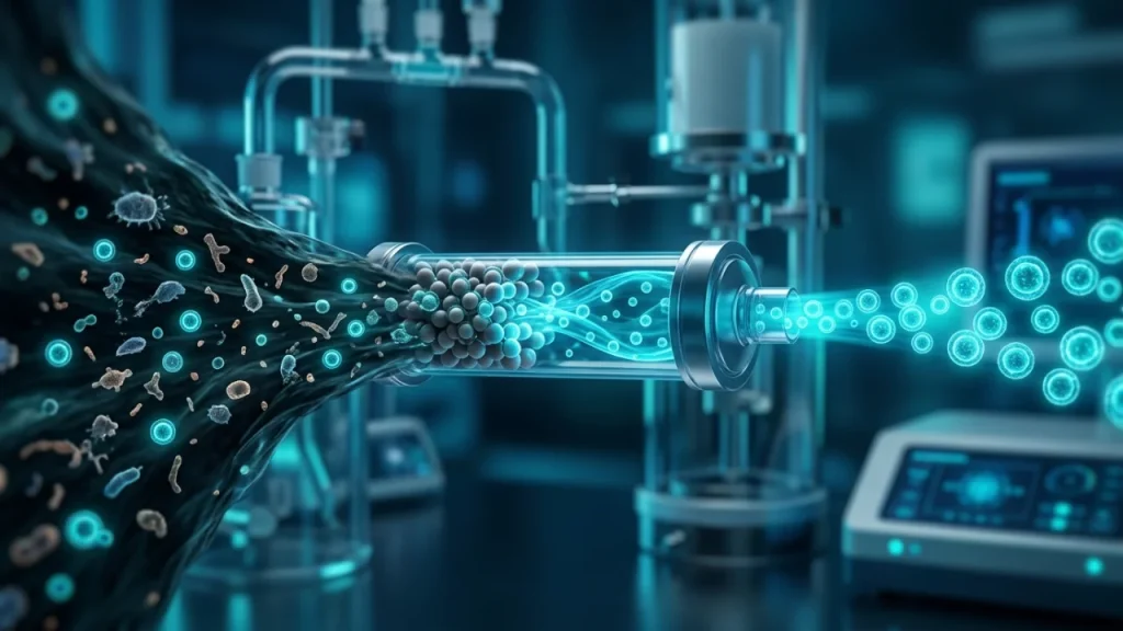
But there is a major challenge. Exosomes from a lab sample are not alone. They are mixed with many other things. Cell debris, proteins, and other vesicles are also present. These contaminants confuse the message. They can cause side effects or reduce treatment power.
That is why purification is not just a step. It is the most critical step. Effective exosomes downstream purification separates the true signal carriers from the noise. Pure exosomes mean predictable, safe, and powerful therapies. Without purity, this brilliant natural system cannot be used reliably in medicine. The next step is understanding how scientists clean up these mixtures to get the useful vesicles alone.
Why Impure Exosomes Can Cause Problems
Impure exosome samples are not just messy. They are potentially dangerous. Contaminants act like unwanted passengers on a delivery truck. They can disrupt the entire mission.
One major problem is false signals. Other vesicles and protein fragments in the mix can send their own messages. Imagine an exosome carrying a “repair” order. A contaminant next to it might send a “stop growth” signal instead. The target cell gets confused. The therapy fails because the instructions conflict.
Safety is the biggest concern. Foreign proteins or cell debris can trigger a strong immune response. The body sees these contaminants as invaders. It launches an attack. This can cause inflammation or serious allergic reactions. A patient could get sick from the treatment itself, not their disease.
Impurities also dilute the power of therapy. If only half the vesicles in a dose are true exosomes, the effect is weaker. Doctors would need to give a larger, impure dose to get the same result. This increases the risk of side effects. It makes treatments unpredictable and hard to study.
Specific contaminants cause specific issues: – Nucleic acid fragments from broken cells can be taken up by target cells. They might interfere with normal cell genes. – Lipids from cell membranes can clog delivery systems or form clumps. – Large protein aggregates can block exosomes from reaching their target.
The goal of exosomes downstream purification is to remove these risks. Pure exosomes give a clear, consistent signal. Their dose can be measured accurately. Their safety profile is reliable. Without this clean separation, clinical trials cannot produce trustworthy results. One batch of therapy might work while another causes harm.
Therefore, purification is not about being neat. It is about ensuring that a revolutionary treatment does not become a source of new problems. The next logical question is how scientists achieve this necessary cleanliness from a complex starting soup.
The Goal of Exosomes Downstream Purification
The goal of exosomes downstream purification is to create a final product with three key qualities. It must be safe, potent, and consistent. Think of it like filtering water from a muddy river. You want only pure water, not dirt, algae, or bacteria.
First, purification aims for safety by removing specific hazards. These include other tiny vesicles that look similar to exosomes. It also removes broken cell fragments and free-floating proteins. Even harmless contaminants are a problem. The immune system may react to them. This reaction could cause fever or inflammation in a patient. A pure sample avoids this false alarm.
Second, the process seeks to concentrate the therapeutic power. A soup full of cell waste dilutes the exosomes. Scientists measure success by the ‘purity ratio’. This ratio compares the number of true exosomes to the total particles in the sample. A high ratio means a strong, reliable dose. A low ratio makes the treatment weak and unpredictable.
Third, purification must protect the exosomes’ natural abilities. Harsh cleaning methods can damage the very structures we need. Exosomes have delicate surface proteins. These proteins act like GPS addresses. They guide the vesicle to the correct target cell in the body. A good purification method keeps this GPS system intact.
How do scientists know they succeeded? They use several checks: – They count particles to ensure a high concentration of exosomes. – They check size to confirm most vesicles are in the correct, tiny range. – They test for marker proteins found only on exosomes. – They look for contaminants like DNA from broken cells.
The final product should be a clear liquid. It should not contain visible clumps or debris. This visual check is simple but important. Achieving all these goals is a balancing act. The method must be aggressive enough to remove impurities. Yet it must be gentle enough to preserve exosome function. This balance is the core challenge of exosomes downstream purification. Mastering it turns a complex cellular soup into a precise medical tool. The next step is to examine the specific methods used to reach this goal.
How Purity Affects Medical Research
Clean exosomes are not just a quality goal. They are a strict requirement for trustworthy science. Imagine a lab studies how cancer exosomes help tumors spread. If their sample is contaminated with free-floating proteins from cell debris, the results will be wrong. Scientists might blame the exosomes for an effect actually caused by the contaminants. This leads to false conclusions and wasted time.
Impure samples create three major problems in research.
First, they hide the true mechanism. Exosomes work through signals on their surface and inside their cargo. Other particles in the soup can send competing signals. Researchers cannot tell which signal caused the observed effect. Was it the exosome, or something else? Purity answers this basic question.
Second, impurities make experiments impossible to repeat. Reproducibility is the foundation of science. Another lab must get the same result using the same methods. If the first lab’s exosomes downstream purification was sloppy, their “exosome” recipe is secret. It contains unknown extras. No one else can copy it exactly. The finding becomes an isolated fact, not a building block for new cures.
Third, contaminants can be toxic to cells in experiments. This skews results dramatically. – Cell growth might slow down because of contaminants, not the exosomes. – Cells might die, suggesting a toxic effect from the exosomes when they are innocent. – Inflammatory responses could be triggered by cell debris, masking the exosomes’ true role.
For example, a study might show exosomes from stem cells heal heart tissue. But if the sample has many broken cell fragments, those fragments could cause inflammation. The healing effect might be weaker or different. Scientists would not get a clear picture of the exosomes’ real power.
Consistent purification solves these issues. It turns unknown mixtures into defined tools. Researchers can then draw accurate maps of how exosomes behave. They can compare studies from different labs with confidence. This collective knowledge accelerates discovery. The path from a lab finding to a patient treatment is long and hard. Clean data from pure exosomes makes that path much straighter and faster. It ensures that every step forward is built on solid ground. The next logical question is how researchers achieve this essential cleanliness in practice.
Common Contaminants in Exosome Samples
Proteins That Stick to Exosomes
Proteins are the most common hitchhikers in exosome samples. They stick to the outside of exosomes like dust on a wet ball. These proteins do not belong to the exosome itself. They come from the fluid cells grew in or from broken cells. Their presence creates a major problem for science.
Think of an exosome as a tiny delivery truck. Its surface has specific address labels. These labels tell it which cells to target. Contaminant proteins are like sticky notes slapped randomly on the truck. They can block the real addresses. They can also send false signals to cells. A cell might react to the sticky note, not the truck.
These extra proteins mainly come from two sources. First, the growth medium that nourishes the parent cells is full of proteins, like albumin. During exosomes downstream purification, these medium proteins can co-pellet with the vesicles. Second, when cells die or get stressed, they spill their contents. This debris includes intracellular proteins and fragments of membranes.
The effects on research are significant and direct. – Diagnostic tests can give false readings. A test might detect a cancer-linked protein. But that protein could be a contaminant, not from the exosome. This leads to wrong conclusions about a patient’s health. – In therapy, contaminant proteins can cause immune reactions. The body might attack the foreign proteins. This could ruin the treatment and harm the patient. – Basic research into exosome function gets muddled. Scientists cannot know if an observed effect comes from the exosome or a clinging protein.
For example, a study might find exosomes that boost inflammation. The real culprit could be inflammatory proteins stuck to them. The exosomes themselves might be harmless or even anti-inflammatory. Without removing these sticky proteins, the true nature of the exosome remains hidden.
Separation is difficult because many contaminant proteins are similar in size or weight to exosomes. They travel together in standard spins and filters. Advanced purification steps are needed to pull them apart. These steps often use antibodies or special chemical tricks to capture either the exosomes or the unwanted proteins.
Cleaning away these protein contaminants is a critical step. It reveals the exosome’s authentic biological identity. This clarity is non-negotiable for turning exosomes into reliable tools. After dealing with protein hitchhikers, scientists must face another tricky impurity: nucleic acids that travel inside.
Nucleic Acids That Hide in Samples
Exosomes do not travel empty. They carry internal cargo, including nucleic acids like RNA and DNA. This is a normal part of their biological role. However, free-floating fragments of these same molecules also exist in the sample fluid. These fragments are a major contaminant. They are not packaged inside exosomes. Yet they can stick to the outside of the vesicles or simply float alongside them. This creates a serious problem for exosomes downstream purification.
During analysis, scientists often break open exosomes to study their genetic contents. The process also releases any contaminating nucleic acids that were hiding in the sample. The test results then show a mix of signals. It becomes impossible to know what truly came from inside the exosome. For instance, a study might find RNA linked to Alzheimer’s disease. That RNA could be from the exosomes. It could also be from dying brain cells in the fluid. The source matters greatly for creating an accurate diagnostic test.
The effects are concrete and disruptive. – Diagnostic errors can occur. A blood test might detect cancer-associated DNA fragments. Those fragments could be free in the blood, not protected inside exosomes. A doctor might wrongly assume the exosomes are from a tumor. – Therapeutic applications face risks. If exosomes are loaded with therapeutic RNA, contaminant RNA could dilute the dose. It could also trigger unintended immune responses. – Basic biology studies get misleading data. Scientists may credit an exosome with carrying specific genetic instructions. The instructions might actually come from contamination.
Separation is tough because these fragments are small. They pass through filters designed to catch larger particles. Specialized methods are required for clean separation. These methods often involve enzymes that digest unprotected nucleic acids. Only the material safely inside exosomes survives. This purification step is non-negotiable for clarity.
After removing proteins and nucleic acids, one major hurdle remains. Scientists must also separate exosomes from other, look-alike vesicles released by cells. This final distinction is crucial for purity.
Other Vesicles That Look Like Exosomes
Cells release a family of tiny bubbles, not just one type. Exosomes are just one member. Other vesicles can look almost identical under a microscope. They are all roughly the same small size. This creates a major purification puzzle.
Think of it like sorting apples. You have two baskets. One holds only Granny Smith apples. The other holds a mix of similar green apples. Telling them apart from the outside is very hard. Scientists face this with vesicles. They must separate the “Granny Smith” exosomes from the other “green apple” vesicles.
The main look-alikes are called microvesicles. They pinch directly off the cell’s outer membrane. This is different from exosomes. Exosomes form inside the cell in special compartments. But once released, they are nearly the same size. Their membranes are also made of similar fats.
Why does this mix-up matter? The cargo inside these vesicles is often different. Their surfaces carry different signals too. – Microvesicles may carry more random cell debris. – They might display different protein markers on their surface. – This affects how they communicate with other cells.
Using a mix for therapy could give unpredictable results. A drug delivery system must be consistent. For accurate research, scientists need to study pure exosomes alone. Contamination skews all the data.
So how do experts tell them apart? They use the vesicles’ unique surface signatures. It is like finding a specific badge on a uniform. Exosomes often carry certain protein markers. Tools like flow cytometry can detect these tags. Another method uses precise density gradients. Different vesicles settle at slightly different levels in a centrifuge tube.
This careful sorting is a key part of exosomes downstream purification. The goal is to get a sample that is only exosomes. This step comes after removing stray proteins and genetic material. It is the final barrier to a clean sample.
Achieving this purity is difficult but essential. The next challenge is confirming what you have actually collected is correct. Verification is its own critical step.
Cell Debris From Broken Cells
When a cell breaks apart, it scatters its contents. This debris is a major contaminant in exosome samples. Think of it like trying to collect only specific letters from a shredded document. You first must filter out the larger confetti.
This debris includes many fragments. They come from broken cell membranes and internal structures. These pieces are often much larger than exosomes. Yet they can carry similar surface proteins. This makes them tricky to separate.
The debris causes several concrete problems for research and therapy:
- It can clog and damage sensitive lab equipment. This includes the fine filters used in early purification steps.
- It provides false signals in experiments. Tests might detect a protein and credit it to an exosome. But that protein could just be floating on a membrane fragment.
- It can trigger unwanted immune reactions if used in a treatment. The body may see the debris as dangerous waste.
The process to remove this material is physical and stepwise. Scientists first use slow, gentle spins in a centrifuge. This is called differential centrifugation. The goal is to pellet the large, heavy debris. The exosomes remain floating in the liquid above.
But this is rarely perfect. Some small debris stays suspended. This is why exosomes downstream purification involves multiple techniques. Filtration through tiny pores is a common next step. These pores are often 200 nanometers or smaller. They let exosomes pass through but block most remaining fragments.
The challenge is that exosomes themselves are only about 30 to 150 nanometers wide. Pushing them through filters can sometimes damage their delicate membranes. It is a careful balance. You must remove contaminants without harming the vesicles you want to keep.
Successful removal of cell debris sets the stage for cleaner later steps. It makes the final isolation of pure exosomes possible. Without this cleanup, the sample is too messy for precise analysis. The next purification stages then face a much harder task. They must sort through a cluttered mixture instead of a refined one. This initial filtering is a foundational act of preparation. It directly impacts the quality and reliability of all subsequent data or therapeutic applications.
Basic Methods for Exosome Isolation
Using Ultracentrifugation to Spin Samples
Ultracentrifugation is the most common method for isolating exosomes. It uses extremely high speeds to create powerful forces. Think of it as a super-powered version of the earlier centrifugation step. The machine, called an ultracentrifuge, spins samples at tremendous velocities. These speeds can exceed 100,000 times the force of gravity.
This force acts like a highly effective sorter. Heavier and denser particles are pushed to the bottom of the tube fastest. Lighter particles settle more slowly or not at all. In a prepared sample, exosomes are among the denser components left. They form a tiny, often invisible, pellet at the tube’s bottom after a long spin.
The process is not just one spin. It is a sequence. A typical protocol involves several steps: – An initial spin at moderate speed to remove any remaining large debris. – A very long, high-speed spin to pellet the exosomes themselves. This can last several hours. – A final wash step. The pellet is resuspended and spun again to remove leftover soluble proteins.
This technique excels at processing large sample volumes. It can concentrate exosomes from liters of fluid down to a small pellet. The method is also versatile. It does not require special chemical tags or filters that might damage membranes.
However, ultracentrifugation has key drawbacks. The intense forces can damage exosomes. Their delicate membranes may rupture or fuse together. The process also co-pellets non-exosome particles with similar density. This includes protein aggregates and other vesicles.
Therefore, the pellet from ultracentrifugation is often not pure. It is an enriched preparation. This outcome highlights why exosomes downstream purification is a multi-stage effort. Ultracentrifugation provides a concentrated batch of vesicles. Yet this batch still contains impurities.
The technique’s success depends heavily on precise settings. Rotor type, speed, time, and temperature are all critical. Small changes can greatly affect the yield and quality of the final pellet. This makes the method powerful but demanding. It requires careful optimization for each sample type.
Ultracentrifugation thus serves as a crucial workhorse in exosome isolation. It delivers a concentrated product for further study. Yet its limitations make clear that isolation is rarely complete after spinning alone. The enriched pellet sets the stage for even finer purification techniques to take over.
Polymer Precipitation for Simple Collection
Polymer precipitation offers a simpler path to collect exosomes. This method uses special water-soluble polymers. These polymers change how molecules dissolve in a liquid. They create a crowded environment in the solution. Exosomes become less soluble under these conditions. They gently fall out of solution as a visible cloud or pellet.
The process is straightforward and does not need complex equipment. A common polymer used for this is polyethylene glycol, or PEG. Researchers mix a concentrated PEG solution with their sample. This could be cell culture fluid or blood plasma. The mixture is then incubated overnight at a cold temperature, often around 4°C. The exosomes slowly gather together during this time.
The final step is a low-speed centrifugation run. This spin is much gentler than ultracentrifugation. It collects the clumped exosomes at the bottom of the tube. The entire protocol can be completed in a standard lab centrifuge. This makes the technique widely accessible.
The key advantage is its simplicity and high yield. It can process many samples at once with minimal hands-on time. The method is also gentle on exosome membranes. It avoids the high shear forces that can damage vesicles during ultracentrifugation.
However, polymer precipitation has significant trade-offs. The major issue is purity. The polymers co-precipitate many other substances alongside exosomes. – Proteins and protein complexes also clump together. – Lipoproteins from blood samples often contaminate the pellet. – Residual polymer can coat the exosomes, potentially affecting later studies.
This means the collected material is often less pure than even a basic ultracentrifugation pellet. The sample requires extensive washing. This washing is a critical part of exosomes downstream purification after precipitation. Without it, the exosome preparation contains too many impurities for reliable research.
The polymer itself can also interfere with certain analyses. It may inhibit downstream steps like RNA sequencing or protein tests. Scientists must often remove all traces of the chemical before they can study the exosomes in detail.
Therefore, this method is excellent for initial collection or diagnostic screening where absolute purity is less critical. It is a powerful tool for quickly concentrating vesicles from large or numerous samples. Yet the output is typically a very crude preparation. It sets a clear stage for rigorous purification methods that must follow. The next techniques must separate the true exosomes from this complex mixture of precipitated material.
Size-Based Filtration With Membranes
Size-based filtration uses membranes with tiny pores. These pores act like a sieve. They physically separate particles by their size. Larger particles get trapped on the filter. Smaller particles pass through. This principle is straightforward and powerful.
For exosome isolation, scientists use filters with very specific pore sizes. A common target is 200 nanometers. This size is key. Most exosomes are smaller than this limit. Many contaminating cells and large debris are bigger. The process often uses multiple filters in a sequence.
A typical workflow might have two main steps. First, a larger pore filter removes big contaminants. Second, a smaller pore filter captures the exosomes themselves. The exosomes are retained on the final membrane’s surface. They can then be washed and collected.
- First, a sample goes through a 0.8 or 0.45 micron pre-filter. This step catches whole cells and large fragments.
- Next, the liquid flows through a 0.22 or 0.1 micron membrane. This captures exosomes and similar-sized vesicles.
- Finally, scientists flush the filter to recover the trapped exosomes in a smaller volume.
This method is fast and does not require complex equipment like ultracentrifuges. It can process several samples at once. The process is also scalable. It can handle large volumes of starting material, like liters of cell culture fluid.
However, filters face clear challenges. The main issue is membrane clogging. Proteins and other sticky substances can block the pores. This slows the flow and can reduce the exosome yield. Pre-filtration helps but does not solve it completely.
Another concern is shear stress. Forcing exosomes through narrow pores under pressure can damage their delicate membranes. This might affect their structure and function. Gentle pressure and specialized membrane materials can lessen this risk.
Purity is another trade-off. Filters separate by size only, not by identity. Anything smaller than the pre-filter but larger than the final pore gets concentrated alongside exosomes. This includes other vesicles and protein aggregates. The result is cleaner than polymer precipitation but not pure.
Therefore, filtration is often an excellent intermediate step. It is highly effective for quick volume reduction and crude isolation. It prepares a sample for more precise techniques that separate by other properties, like density or surface markers. This sets the stage for advanced purification methods needed for rigorous research.
Limits of These Basic Techniques
Basic isolation methods get exosomes out of solution, but they rarely get *only* exosomes. This is their core limit. They co-isolate other particles that share a simple physical trait, like size or solubility. These unwanted hitchhikers are called contaminants. They can severely muddy research results or ruin a therapeutic batch.
Think of it like fishing with a wide net. You want salmon. Your net catches all medium-to-large fish. You will also get trout, bass, and other species of similar size. Basic methods work the same way. They lack the precision to pick only exosomes from the cellular debris.
The main contaminants fall into a few key groups. First are other extracellular vesicles. Microvesicles are often larger than exosomes but can break into smaller pieces. Apoptotic bodies from dying cells are much larger, yet fragments remain. These vesicles have different functions and contents than exosomes.
Second are non-vesicular particles. Lipoproteins like HDL and LDL are common in blood samples. Protein aggregates clump together to mimic vesicle size. These aggregates lack a membrane and carry no genetic cargo, but they are often present.
Third is soluble junk. Leftover growth factors and free-floating proteins like albumin persist. They stick to the outside of exosomes or form complexes. Nucleic acids from broken cells can also attach to vesicle surfaces.
These contaminants create major problems for scientists. They distort protein analysis. A detected signal might come from a contaminant, not the exosome. They skew genetic studies. RNA found could be from outside, not packaged inside the vesicle. In therapy, contaminants could trigger immune reactions or block the exosome’s intended target.
Each basic method has a specific contamination profile. Ultracentrifugation pellets everything of a certain density together. Polymer precipitation brings down exosomes but also precipitates other molecules. Size-based filtration concentrates anything that fits through the pores.
This is why exosomes downstream purification is not a luxury. It is a necessity for quality science and safe medicine. Basic isolation is just the first crude step. The real challenge starts after this step: refining the harvest into a pure, defined population of exosomes. This requires more sophisticated tools that can tell exosomes apart from their look-alikes based on finer details than just size or weight.
Advanced Exosomes Downstream Purification Techniques
How Size-Exclusion Chromatography Works
Size-Exclusion Chromatography is a gentle and precise filtering method. It does not crush or force particles through a membrane. Instead, it uses a column packed with tiny porous beads. These beads are like sponges with many tunnels inside.
The mixture of exosomes and contaminants is added to the top of the column. A liquid buffer then washes everything down through the bead bed. This is where the magic happens. The separation depends on how deeply each particle can enter the bead pores.
Small contaminants like proteins and free RNA are very tiny. They wiggle deep into the bead tunnels. This takes them on a long, winding path through the column. Their journey is slow.
Exosomes are much larger than simple proteins. They are too big to enter most pores. So they cannot take those winding internal paths. The exosomes flow around the outside of the beads instead. This route is faster and more direct.
The largest particles, like cell debris or big protein clumps, are also excluded. They take the fast path around the beads. This is why exosomes downstream purification often uses SEC in combination with other methods for the cleanest results.
The liquid coming out the bottom of the column is collected in small tubes called fractions. The order of what comes out is predictable. First, you collect the large particles that took the fast path. Then, you collect the pure exosomes. Finally, the small contaminants that got stuck in the beads slowly emerge.
This process gives scientists a clear separation based strictly on size. It is excellent for removing small soluble junk. The exosomes stay intact and functional because there is no harsh force applied. However, SEC alone may not separate exosomes from other vesicles of identical size, like some lipoproteins.
The result is a cleaner sample ready for precise analysis or therapeutic use. The next step often involves looking at surface markers to confirm identity after this size-based clean-up.
Tangential Flow Filtration for Large Volumes
Processing liters of cell culture fluid for exosomes is a major challenge. Simple filters clog almost instantly. Tangential Flow Filtration solves this problem with a clever design. The fluid flows *across* the filter surface, not straight through it.
This sideways flow sweeps away particles that might block the membrane. It keeps the filter clean and working. The process uses special membranes with precise pore sizes. These pores are measured in nanometers, which are billionths of a meter.
Small molecules like salts and sugars pass through the pores. They are removed as waste fluid. Larger exosomes cannot fit through the tiny pores. They are retained and become more concentrated over time. This method is highly efficient for exosomes downstream purification from big volumes.
The system offers key advantages for scaling up work. – It can process volumes from milliliters to hundreds of liters. – The gentle flow protects delicate exosome structures. – It works as both a concentration and a purification step.
Scientists often choose a membrane with a 100-200 nanometer pore size. This size captures most exosomes while letting smaller contaminants escape. The process is continuous and can be automated. This makes it ideal for preparing large, clean samples for further study.
After TFF, the exosome sample is much smaller in volume. It is also cleaner from small soluble waste. However, it may still contain particles similar in size to exosomes. This is why TFF is typically a first major concentration step.
Later techniques, like the size-exclusion chromatography discussed earlier, provide final polishing. TFF handles the heavy lifting of volume reduction. This combination is powerful for industrial and clinical applications needing vast amounts of pure exosomes.
The method proves essential when moving from lab research to therapeutic production. It turns an impossible task into a manageable routine. Next, we will explore techniques that separate exosomes based on their unique surface features, adding another layer of precision.
Affinity Capture Using Specific Bonds
Affinity capture isolates exosomes with pinpoint accuracy. This method uses molecular “keys” to find specific “locks” on an exosome’s surface. It targets proteins or markers unique to certain exosome types. This approach provides exceptional purity for demanding applications.
The process relies on known biological interactions. Antibodies are common tools. They are proteins designed to bind one specific target molecule. Scientists attach these antibodies to solid surfaces like beads or plates.
Exosomes flow over these antibody-coated surfaces. Only exosomes carrying the correct surface marker stick. Everything else washes away. This is a powerful form of exosomes downstream purification. It yields a sample defined by its biology, not just its size.
Several capture strategies are widely used. – Antibody-based capture targets proteins like CD63, CD9, or CD81. These are common on many exosomes. – Lectin-based capture binds to specific sugar groups on the exosome membrane. – Tag-based capture uses engineered exosomes. The cells producing them are modified to add a special “tag” protein. This tag is then easily captured.
Each strategy has clear pros and cons. Antibody methods offer high specificity but can be costly. Lectin methods are cheaper but may be less specific. Tag-based methods give superb purity but require genetic engineering of the source cells first.
This technique is crucial for clinical research. Imagine studying exosomes from cancer patients. You need to isolate only those from tumor cells. Affinity capture can target a cancer-specific marker. This lets researchers analyze signals just from diseased cells.
The process is gentle on exosome structure. It does not involve harsh forces or pressure changes. Captured exosomes can often be released later for functional studies. A mild change in acidity or a special solution breaks the bond.
However, affinity capture has limitations. It requires prior knowledge of the target marker. Not all exosome subtypes have a known unique signature. The binding step can also be slow compared to filtration methods.
This method is rarely used alone in large-scale workflows. It typically follows initial concentration steps like TFF. The combined process is powerful. First, reduce the volume and remove bulk contaminants. Then, use affinity capture to pick out the exact exosome population you need.
Such precision is vital for creating reliable diagnostics and targeted therapies. The next logical step is analyzing what this pure cargo contains, moving from isolation to characterization.
Combining Methods for Better Purity
No single technique can isolate perfectly pure exosomes in one step. Each method removes certain contaminants but leaves others behind. The solution is a smart sequence of steps. This staged approach is called exosomes downstream purification.
Think of it like filtering water. You might use a coarse sieve first to remove leaves. Next, a fine cloth catches sand. Finally, a carbon filter takes out chemicals. Similarly, exosome purification uses broad methods first, then precise ones.
A common workflow starts with ultrafiltration or TFF. This quickly concentrates the sample. It removes most of the free proteins and small molecules. The volume shrinks dramatically. This prepares the sample for more delicate steps.
The next step often involves size-based separation. Size-exclusion chromatography is a frequent choice here. It further separates exosomes from similar-sized particles. This step polishes the sample, enhancing purity.
Finally, an affinity-based method can select a specific exosome group. This targets a surface marker unique to the exosomes of interest. The result is a highly purified population ready for analysis or therapy.
Combining methods solves individual weaknesses. For example: – Ultracentrifugation is good for large volumes but can damage exosomes. Following it with gentle size-exclusion chromatography can help repair the sample’s quality. – Precipitation methods are simple but co-precipitate many impurities. Running the result through a size column effectively cleans it up. – Tangential flow filtration is excellent for concentration but not perfect separation. Adding a polishing step like chromatography creates a superior final product.
This multi-step process is essential for reliable research. Contaminants can skew data and lead to false conclusions. Pure exosomes ensure scientists study the real signals.
The exact combination depends on the starting material and goal. Exosomes from blood need different steps than those from cell culture. Diagnostic applications may prioritize speed, while therapeutic ones demand extreme purity.
Each added step increases purity but can lower yield. Some exosomes are lost at each stage. Scientists must balance this trade-off for their specific needs.
Advanced workflows are becoming more standardized. This consistency lets labs worldwide compare their results with confidence. It turns exosome science from an art into a more precise engineering discipline.
The final output of this careful process is a pristine exosome preparation. These vesicles have their native structure and cargo intact. They are now ready for the next critical phase: detailed analysis to unlock their secrets.
Step-by-Step Purification Process
Starting With Clean Sample Preparation
The quality of your final exosome sample is decided in the very first steps. Everything you do at the start either helps or hurts the later purification stages. Think of it like cooking. If you begin with spoiled ingredients, even the best techniques cannot save the meal. The same is true for exosome research. A messy starting material makes clean results almost impossible.
Cells grown in a lab dish are a common source. Scientists call this cell culture. The fluid around these cells holds the exosomes. This fluid must be handled with care from the moment it is collected. It should be processed quickly or frozen in a specific way. Slow handling lets cells break apart and release extra debris. This debris will later contaminate the exosome pool.
Blood and other bodily fluids are trickier starting points. They contain many more components that can interfere. For example, blood has lipoproteins. These particles are similar in size to exosomes. They can easily be mistaken for exosomes in later tests. Other fluids like urine or saliva have their own unique contaminants. Each source requires a tailored approach right from the beginning.
Good sample preparation follows several key rules. First, use gentle centrifugation steps early on. This removes whole cells and large cell fragments. Second, avoid repeated freezing and thawing of samples. This process can break exosomes apart. Third, filter the sample through a fine pore filter. This removes even more unwanted large particles before the main purification begins.
These careful initial steps set the stage for effective exosomes downstream purification. They remove the biggest sources of contamination upfront. This makes the job of later techniques like chromatography much easier. A clean start improves both the purity and the yield of the final exosome preparation. Scientists spend less time fighting impurities and more time studying the exosomes themselves.
Ignoring this phase creates major problems. Contaminants can clog expensive filtration equipment. They can also bind to chromatography columns, ruining them for future use. Most importantly, they lead to wasted time and resources. A purification run that starts with a poorly prepared sample often ends with failure. The entire process must be repeated.
Therefore, successful exosome science demands respect for the preparation stage. It is the unseen but critical work that makes all advanced analysis possible. By investing time here, researchers ensure their downstream data reflects true biology, not experimental artifacts. This foundational discipline turns raw biological material into a reliable scientific resource ready for precise characterization.
Removing Large Debris First
The first major step in exosomes downstream purification is often a spin in a centrifuge. This machine spins samples at high speeds. It uses centrifugal force to separate particles by their size and weight. Larger, heavier particles sink to the bottom faster than small, light ones. Think of it like a whirlpool. Heavy rocks sink immediately. Fine sand takes much longer to settle.
Scientists use a series of controlled spins at increasing speeds. Each spin targets a different type of contaminant. The goal is to remove everything bigger than an exosome first. This clears the path for later, more precise techniques.
A standard initial series involves three main spins: – A low-speed spin at around 300 times gravity for ten minutes. This pellets whole cells and very large debris. – A medium-speed spin at about 2,000 times gravity for twenty minutes. This removes smaller cell fragments and large organelles. – A final higher-speed spin at 10,000 times gravity for thirty minutes. This pellets even smaller vesicles and protein aggregates that are still larger than exosomes.
After each spin, the researcher carefully collects the supernatant. The supernatant is the liquid on top of the solid pellet. The unwanted material forms the pellet at the tube’s bottom. The supernatant, now cleaner, is moved to a new tube for the next step. The pellets of debris are discarded.
This process is foundational but has limits. Centrifugation alone cannot isolate pure exosomes. It is a bulk separation method. Some particles similar in size to exosomes may remain. Furthermore, very high forces can sometimes damage the exosomes themselves if applied for too long. That is why this step is carefully calibrated.
The success of this stage directly impacts everything that follows. A well-cleared supernatant prevents clogging in ultrafiltration devices. It also protects sensitive chromatography columns from fouling. Effective removal of large debris means subsequent steps can focus on finer separations. This increases the final yield of intact exosomes.
In essence, this sequential centrifugation is a coarse filter. It does the heavy lifting of gross purification. It prepares the sample for the fine-tuning of later methods like ultracentrifugation or size-exclusion chromatography. Without this clear removal of large debris, the entire downstream process becomes inefficient and unreliable.
Concentrating Exosome Solutions
After removing large debris, the exosome sample is still very dilute. It exists in a large volume of liquid. This makes direct analysis or further purification difficult. Think of it like searching for a specific fish in a huge, cloudy lake. Draining most of the water would make the fish much easier to find and catch. Concentration does exactly that for exosomes.
The goal is to reduce the sample volume. This increases the number of exosomes per drop. A concentrated sample is essential for all exosomes downstream purification and analysis steps. It improves the sensitivity of detection methods. It also makes subsequent separation techniques far more efficient.
Scientists use several key methods to achieve this concentration. Each method has different principles and advantages.
- Ultrafiltration uses membranes with tiny pores. These pores allow water and small molecules to pass through under pressure. The larger exosomes cannot fit through the pores. They are retained and become concentrated in a smaller volume. This method is relatively fast and scalable.
- Precipitation involves adding special polymers or salts to the sample. These chemicals change the solution’s properties. They make exosomes less soluble in the liquid. The exosomes then clump together and fall out of solution as a pellet. This pellet can be collected after a low-speed spin.
- Tangential Flow Filtration is a gentler, continuous process. The sample flows across a membrane, not directly into it. This parallel flow reduces clogging and shear stress. It is excellent for processing larger volumes while keeping exosomes intact.
Choosing the right method depends on the sample’s starting volume and the need to preserve exosome quality. Harsh methods can damage the fragile vesicle structure. They can cause exosomes to rupture or stick together irreversibly. This would ruin their natural function as delivery vehicles.
A successful concentration step yields a potent, small-volume sample. This robust preparation is now ready for the final, precise isolation stages. The path to pure exosomes becomes clearer after this crucial volume reduction.
Final Polishing for Highest Purity
The concentrated sample still contains impurities similar in size to exosomes. These include protein aggregates and other tiny vesicles. Final polishing steps remove these leftovers. This stage is critical for research and therapy. Pure exosomes give clear, reliable results.
Scientists often use density gradients for this final clean-up. They layer the sample onto a special solution. This solution has a density that increases from top to bottom. The tube is then spun at very high speeds in an ultracentrifuge.
Each particle in the sample migrates to its matching density zone during the spin. Exosomes settle at a specific density range. Most contaminants float to other levels. This physically separates them based on buoyancy. The exosome band can then be carefully extracted with a pipette.
Size-exclusion chromatography is another key polishing tool. The sample is passed through a column packed with porous beads. Smaller molecules and proteins get trapped in the bead pores. They travel slowly through the column.
Larger exosomes flow around the beads. They move faster and elute first. This method separates particles by their hydrodynamic size. It is gentle and preserves exosome structure and function well. The process is sometimes called exosomes downstream purification.
These high-resolution methods solve specific problems. Density gradients excel at separating vesicles of similar size but different makeup. Chromatography is superb for removing contaminating proteins and small molecules. The choice depends on the primary impurity target.
The final steps have important trade-offs. They yield extremely high purity but can reduce overall quantity. Some exosomes may be lost during handling. The processes also require specialized equipment and more time.
Successful polishing results in a pristine exosome preparation. This sample is now ready for detailed analysis or therapeutic loading. Researchers can confidently study its contents and effects. Therapists can trust its consistency.
This concludes the step-by-step purification journey. From raw fluid to pure vesicles, each stage has a clear goal. The next logical phase involves analyzing what these purified exosomes contain and how they function.
Tools and Equipment for Purification
Choosing the Right Centrifuge
The force of a centrifuge is the engine of exosome isolation. It separates particles based on their size and density. Spinning pushes heavier parts to the bottom of the tube. Lighter parts stay closer to the top.
This force is measured as Relative Centrifugal Force (RCF). You may also see it called “g-force.” It is not just about rotor speed in revolutions per minute (RPM). RCF accounts for the rotor’s radius. A higher RCF means a stronger separating push.
Choosing the correct RCF and time is a balancing act. The goal is to pellet the exosomes you want. You must also leave unwanted debris behind.
First, low-speed spins clear out big junk. Cells and large cell fragments are heavy. A short spin at 2,000 x g for 30 minutes will pellet them. The exosomes remain floating in the liquid above, called the supernatant.
Next, a medium-speed spin removes smaller nuisances. Apoptotic bodies and other large vesicles come down now. A typical step uses 10,000 x g for 45 minutes. This further cleans the supernatant.
The final ultracentrifugation step pellets the exosomes themselves. This requires very high force. Speeds often exceed 100,000 x g. The spin can last over 70 minutes.
The exosomes are tiny but dense enough to form a pellet. The right combination of speed and time is key. Too little force or time leaves exosomes in solution. Too much can crush them or pack them too tightly.
Think of it like filtering different sizes of sand. A gentle shake settles only the biggest rocks. A harder, longer shake gets the finest grains.
Key factors influence your centrifuge choice: – Maximum RCF: Can it reach the needed force? – Rotor type: Fixed-angle rotors are common. Swinging-bucket rotors are gentler. – Capacity: How many samples can you process at once? – Cooling: Heat can damage exosomes. A refrigerated centrifuge is vital for long spins.
This careful progression of spins is part of effective exosomes downstream purification. Each step uses specific tools to remove a class of contaminants. Mastering these settings ensures a cleaner starting material for later polishing stages. The centrifuge, therefore, sets the stage for all subsequent refinement.
Filters and Membranes for Different Sizes
After centrifugation, you have a mix of particles. Exosomes float with similar-sized contaminants. Filtration solves this by using precise physical barriers. These barriers are membranes with tiny holes, or pores.
Pore size is measured in nanometers. One nanometer is one-billionth of a meter. Exosomes typically range from 30 to 150 nanometers in diameter. A filter’s pores must be smaller than unwanted debris but larger than the exosomes. Or the opposite is true, depending on the goal.
Two main strategies exist. The first is dead-end filtration. The liquid is pushed straight through a membrane. Larger particles get stuck on top. Smaller exosomes pass through. This is good for quick clarification.
The second strategy is tangential flow filtration. Here, liquid flows across the membrane’s surface. Pressure forces small molecules through the pores. Larger particles like exosomes stay behind. This method is gentler. It avoids clogging the filter quickly.
Choosing the right pore size is critical. A 200 nm filter will remove large microvesicles and debris. A 100 nm filter can concentrate an exosome sample. It allows smaller proteins and lipids to wash away.
Filters are not perfect. Some challenges exist. – Membrane adsorption: Exosomes can stick to the filter material. – Shear stress: High pressure can damage delicate exosome membranes. – Clogging: Too much debris blocks pores and stops the flow.
These hurdles are part of advanced exosomes downstream purification. Scientists often use filters in series. They might start with a 0.8 micron pre-filter. Then they use a 0.2 micron filter. Finally, a 100 nm membrane captures the exosomes.
The material of the membrane also matters. Polyethersulfone is common. It has low protein binding. Cellulose membranes are another option. They are often used for their gentle properties.
This process delivers a cleaner sample. It removes contaminants that centrifugation might miss. Filtration acts as a precise sieve. It prepares exosomes for the final stages of analysis or therapeutic loading.
The next step often involves capturing exosomes by their surface markers. Filtration provides the clean slate needed for that precision work.
Chromatography Systems Explained
Chromatography is a powerful separation method. It sorts molecules by their physical and chemical traits. Think of it as a race through an obstacle course. Different particles move at different speeds. This separates them cleanly.
For exosomes, liquid chromatography is key. A liquid buffer carries the sample through a column. This column is packed with special beads. These beads act as the obstacles in the race. Exosomes interact with the beads. Contaminants interact differently. This causes separation.
Two main forces drive this process. One is size exclusion. The other is affinity binding. Size exclusion chromatography uses beads with tiny pores. Small molecules enter these pores and get delayed. Larger exosomes cannot enter the pores. They flow around the beads and exit the column first. This method is gentle. It does not rely on strong binding forces.
Affinity chromatography is more specific. The beads are coated with capture molecules. These molecules stick to targets on the exosome surface. Common targets include CD63 or CD81 proteins. The exosomes bind to the beads. Everything else washes away. Then scientists change the buffer solution. This releases the pure, captured exosomes.
The equipment system has several core parts. – A pump creates a steady flow of liquid. – An injector introduces the sample into the stream. – The column is where separation happens. – A detector monitors what comes out. – A fraction collector gathers the purified samples.
This process offers high resolution. It can separate exosomes from similar-sized particles. It also handles larger sample volumes well. This makes it vital for exosomes downstream purification. The goal is a highly concentrated and clean final product.
Scientists choose the method based on their needs. Size exclusion gives a fast, gentle separation. Affinity chromatography provides extreme purity. Sometimes, methods are used in sequence. Filtration might come first to remove debris. Then chromatography refines the sample further.
The result is a preparation ready for detailed analysis or therapeutic loading. Chromatography adds a layer of precision that simpler methods cannot match. It isolates exosomes based on their inherent biological identity. This precise isolation is crucial for reliable research and safe medicines.
Next, we will look at how scientists verify the success of this entire purification process. They need to check what they have actually collected.
Lab Setup for Safe Handling
A single speck of dust can ruin months of work with exosomes. These tiny vesicles are easily contaminated by bacteria, foreign proteins, or even other particles from the air. Contamination destroys sample purity. It makes experimental results unreliable. For therapies, it could be dangerous. Therefore, the lab space itself is a critical piece of equipment. Scientists perform exosomes downstream purification in controlled, clean environments.
The most important tool is the biosafety cabinet. This is a sealed workspace with a glass front. A constant stream of filtered air flows inside it. This air flow pushes contaminants away from the sample. It also protects the scientist. Researchers do all open-container work here. This includes steps like loading samples into chromatography systems or preparing buffers. There are different cabinet classes. The choice depends on the source of the exosomes.
General lab cleanliness follows strict rules. – Surfaces are wiped with special solutions before and after work. – Researchers wear gloves, lab coats, and sometimes face masks. – All tubes and liquids used are sterile. They are often opened only inside the cabinet. – Personal items like phones or notebooks stay outside the clean zone.
Temperature control is another key factor. Exosomes are sensitive to heat. Many purification steps are done in a cold room or using chilled equipment. This slows down degradation. It keeps the exosomes stable. For long-term storage, pure exosome samples go into ultra-cold freezers. These can reach minus eighty degrees Celsius.
The goal is to create a seamless chain of cleanliness. The exosomes move from the biosafety cabinet to a purified state without ever touching unclean air or surfaces. This protects the investment of time and effort in the complex purification process. A single lapse can undo the precision of chromatography or filtration. Consistent, careful handling ensures that the final product is truly pure and useful. Next, we will examine how scientists prove their purification was a success by analyzing what they collected.
Quality Control in Exosomes Downstream Purification
Testing Purity With Protein Assays
After the careful purification process, scientists must answer a critical question. Did it work? They need to test the purity of their collected exosomes. One major test looks for leftover proteins. These proteins should not be there in a pure sample.
Think of exosome purification like filtering sand from a gold nugget. The gold is the exosome. The sand represents common protein contaminants. Even after filtering, some tiny sand grains might stick to the nugget. Protein assays find that leftover “sand.”
Why are proteins a key marker? Cell culture fluid is full of them. They are not part of the exosomes themselves. They float freely in the solution. A successful exosomes downstream purification process removes these free proteins. Their presence means the purification was not complete.
Scientists use several methods to detect these proteins. Each method has a different strategy.
- The BCA assay uses a color change. Proteins interact with copper ions. This turns a solution from green to purple. The deeper the purple, the more protein is present.
- The Bradford assay also uses color. A blue dye sticks to proteins. The solution turns from brown to blue. More blue means more protein.
- UV absorbance is simpler. Proteins absorb light at 280 nanometers. A machine shines light through the sample. Less light passing through suggests more protein.
Researchers often run these tests at two points. They test the original, messy cell culture fluid first. They test the final, purified exosome sample second. Comparing the two results shows how much protein was removed. A large drop is a good sign.
The goal is not always zero protein. Some proteins belong on the exosome’s surface. The assays measure total protein. Very low levels suggest the sample is mostly clean exosomes. High levels are a red flag. They show that free contaminants remain.
This quality control step is non-negotiable. It provides hard numbers on purity. Without it, researchers cannot trust their samples. Contaminated exosomes could ruin an experiment or a therapy. Protein testing acts as a final check on the entire purification workflow. It confirms that the clean handling and complex separation steps paid off. Next, we will look at another crucial test: counting the exosomes themselves to measure yield.
Checking for Nucleic Acid Contamination
Exosomes can carry valuable genetic cargo inside them. This cargo includes RNA molecules with instructions for cells. However, free-floating DNA and RNA outside the exosomes are contaminants. These impurities must be measured and removed. This process is a key part of exosomes downstream purification.
Why is this check so important? Free nucleic acids can cause major problems. They can trigger false signals in diagnostic tests. In therapies, they might cause unwanted immune reactions. Scientists need to confirm that any genetic material in the sample is safely packaged inside exosomes. It must not be loose in the solution.
Researchers use specific tools to find these impurities. The methods look for the unique chemical structure of DNA and RNA.
- Fluorescent dyes are a common tool. These dyes glow when they stick to nucleic acids. They do not glow when they touch exosome lipids or proteins. Scientists mix a dye with the sample. The strength of the glow shows how much free DNA or RNA is present.
- PCR-based methods are extremely sensitive. They can find tiny traces of contaminant DNA. This technique copies a specific genetic sequence over and over. If contaminant DNA is present, the machine will detect the copying process. A positive signal means the sample is not clean.
- Gel electrophoresis is a visual check. Scientists load the sample onto a jelly-like slab. An electric current pulls nucleic acids through the gel. Contaminants form visible bands. Pure exosome samples should show no bands.
A clean result depends on the exosome source. Exosomes from blood plasma often have more background nucleic acids. Those from cell culture can have less. The purification process must remove these materials effectively.
Testing happens after the main isolation steps. Scientists compare the purified exosomes to the starting material. A large drop in detectable free nucleic acids means the process worked. This verification is essential for reliable research. It also matters for creating safe therapies. The next step is ensuring the exosomes themselves are intact and functional.
Using Microscopy to See Particles
Microscopy lets scientists see exosomes directly. This is a crucial visual check. It happens after the chemical tests for purity. Scientists need to confirm the particles are intact. They also need to see their size and shape.
Two main types of microscope are used for this job. Each one offers a different view.
- Transmission Electron Microscopy, or TEM, provides highly detailed pictures. It uses a beam of electrons instead of light. Exosomes are prepared on a small grid. They are stained with heavy metals like uranium. This stain helps outline their shape. The resulting images look like dark, cup-shaped circles. TEM can show if exosomes are broken or have clumped together. It is a key tool for exosomes downstream purification quality control.
- Cryo-Electron Microscopy is a more advanced technique. Samples are frozen very quickly into a glass-like state. This preserves their natural shape perfectly. Scientists can then see exosomes in their native, rounded form. They are not flattened or distorted. Cryo-EM can also show tiny structures on the exosome surface.
Seeing the particles confirms the isolation worked. A good sample will show many separate vesicles of similar size. They should not be empty shells. They should not be large blobs of cellular debris.
The images give specific data. Scientists can measure particle diameter directly from the photos. They can count particles in a field of view. This helps estimate concentration. Most importantly, they check for damage. Broken exosomes will not work as effective delivery systems.
This visual proof complements the nucleic acid tests. A sample can be chemically clean but physically ruined. Microscopy catches this problem. It ensures the vesicles survived the purification process intact.
After confirming physical structure, the next question is about function. Do these intact exosomes still perform their natural biological roles? The next checks will test their activity and contents.
Ensuring Batch-to-Batch Consistency
Producing one perfect batch of exosomes is a scientific achievement. Producing identical batches every single time is a medical necessity. This is the core challenge of exosomes downstream purification. Treatments require predictable performance. A patient’s first dose must match their tenth dose in purity and power.
Think of it like a medicine factory. Every pill must have the same amount of the active drug. For exosome therapies, the “active drug” is the vesicles themselves. Their quality defines the treatment. Inconsistent batches are unsafe. They can lead to unreliable results in patients.
Several factors threaten consistency from one run to the next. The starting biological material can vary. Cells might be from different passages or donors. Their growth conditions can shift slightly. The purification process itself must be tightly controlled. Even small changes in temperature or timing can alter the final product.
Scientists use strict protocols to fight this variation. They define every step with precision. This creates a standard operating procedure. The goal is to remove guesswork and human error.
Key parameters are tracked for each batch. This data forms a consistency profile. – Particle concentration must fall within a target range. – The average size distribution should be nearly identical. – Critical markers on the exosome surface need to be present at stable levels. – The amount of contaminating protein must stay extremely low.
Advanced analytics are essential here. Tools like nanoparticle tracking analysis give detailed size and count data. Flow cytometry checks for surface markers. These tools generate hard numbers. Scientists compare these numbers across batches. They look for statistical outliers.
A successful process shows a tight cluster of data points. All batches land close to the target values. This proves the purification pipeline is robust. It is not a fragile art but a reliable engineering feat.
Documenting everything is crucial. Scientists keep detailed records of every material and action. This is called traceability. If a problem arises, they can trace it back to its source. They can then adjust the protocol to prevent it from happening again.
Ultimately, batch-to-batch consistency builds trust. It assures regulators that the therapy is controllable. It gives doctors confidence in the treatment’s effects. For patients, it means safety and predictable care. The final step before clinical use is confirming this reliability through rigorous, repeated testing.
Scaling Up for Clinical Production
Moving From Lab Scale to Large Volumes
Scaling up exosome production is a major engineering task. Making a few milliliters in a lab is very different from making liters for a clinic. The core challenge is maintaining purity and function at a much larger size. Simple methods do not work well at big scales.
Consider cell culture. In a lab, cells grow in small flasks. For large volumes, scientists use big bioreactors. These are like giant, computerized tanks. They constantly monitor conditions. They control temperature, oxygen, and nutrient levels. The goal is to keep millions of cells healthy and productive. Cells must release the same high-quality exosomes as they did in small flasks.
The harvest step also changes. Instead of handling a few bottles, technicians process hundreds of liters of fluid. This fluid contains the exosomes mixed with many other things. It contains cell debris, proteins, and other waste. The initial concentration of this large volume is a key first step. Scientists often use a method called tangential flow filtration for this. It gently concentrates the fluid without damaging the delicate exosomes.
The exosomes downstream purification process faces the biggest changes. Lab-scale techniques are often too slow or inefficient for big volumes. They cannot handle the large amount of material. Scientists must adapt their purification strategy.
- Centrifugation steps may need longer times or different equipment.
- Chromatography columns must be much larger to capture more exosomes.
- Filtration systems require bigger surface areas to process fluid faster.
Each change introduces new variables. A filter that works for a small sample might clog quickly with a large one. The forces in a large centrifuge can differ from a small one. Scientists must test each scaled-up step carefully. They must ensure it still removes contaminants effectively. The final product must still meet all the strict quality targets set during process development.
The entire workflow needs rethinking for flow and time. At lab scale, a process might take two days. At clinical scale, efficiency is critical. Scientists design the system to minimize delays between steps. This helps keep the exosomes stable and active.
Ultimately, scaling up is about smart engineering. It applies consistent scientific principles to much bigger equipment. The next phase after this is ensuring these large batches remain sterile and safe for patient use.
Automating Purification Steps
Automation brings a new level of control to exosomes downstream purification. Human hands are excellent for small experiments. They are not ideal for making thousands of identical medicine doses. Machines can perform the same action perfectly every single time. This repeatability is vital for clinical production.
Think about a centrifugation step. A scientist might manually set a timer. They might load tubes one by one. An automated system handles all of this. It precisely controls the speed and the duration. It moves samples without pause. This removes small differences between batches. Every batch gets the exact same treatment.
The same principle applies to filtration and chromatography. These are core purification steps. Automated systems manage the fluid flow and pressure. They can detect if a filter is starting to clog. The system can then adjust parameters or alert an engineer. This protects the exosomes from damage. It also ensures contaminants are removed consistently.
Automation directly reduces major risks. Human error is a big source of problems in complex processes. A missed step or a mislabeled sample can ruin an entire batch. Machines follow their programmed protocol without distraction. They also create a detailed digital record. Every action is logged with a timestamp. This provides perfect traceability.
Here is what an automated purification line might handle: – Moving fluids between different purification modules without exposure to air. – Taking small samples for instant quality checks during the run. – Cleaning equipment thoroughly between batches to prevent cross-contamination.
The goal is a “closed system.” Exosomes flow from one step to the next inside sealed tubing. They do not touch the outside environment. This is almost impossible to do reliably by hand at a large scale. Automation makes it possible. It safeguards both the product and the patient.
This shift requires careful planning. Engineers and scientists must translate their manual methods into code and machine instructions. They test the automated process extensively. They prove it works as well as the manual method, but with far greater consistency. The result is a robust pipeline ready for making medicine.
Automation turns a scaled-up process from a challenging manual operation into a reliable factory. The next focus becomes monitoring the output of this factory to guarantee every vial meets the strictest standards.
Meeting Regulatory Standards for Safety
Every medicine intended for human use must meet strict government rules. These rules exist for one primary reason: patient safety. They ensure that a therapy is both safe and effective. Exosome therapies are no exception. The path from lab to clinic is governed by these standards.
Think of it like a recipe for a life-saving treatment. The recipe must be followed exactly every single time. Regulatory standards define that perfect recipe. They cover the entire production process. This includes every step of exosomes downstream purification. The rules specify how pure the final exosomes must be. They set limits for any leftover cell debris or chemicals from the process.
A key concept is “characterization.” Scientists must prove they know exactly what is in their exosome product. They must answer several critical questions. – What specific molecules are on the exosome surface? – How many exosomes are in each dose? – Are any dangerous contaminants present, even in tiny amounts?
This requires powerful tools and constant testing. Scientists use machines to count particles and analyze proteins. They check for bacterial or viral contamination. All this data is collected in a massive file called a regulatory submission.
Consistency is another major rule. Regulators do not approve a single perfect batch. They approve the *process* that makes the medicine. They need proof that the process works the same way every time. This is where automation from the previous section becomes vital. A machine-driven process provides the consistent data regulators demand.
The rules also demand rigorous tracking. This is known as traceability. If a single vial of exosomes is given to a patient, its entire history must be known. Which donor cells did it come from? On what date was it purified? What were the test results? This chain of information is crucial for safety.
Meeting these standards takes years of careful work. Scientists must design their studies to answer regulatory questions from the start. The cost of failure is high. A process that cannot meet these standards will never help patients.
Ultimately, these regulations transform exosomes from a research tool into a trusted medicine. They provide the blueprint for quality that doctors and patients can rely on. The next step is using this high-quality product in precise ways to treat disease.
Cost Considerations in Scaling
Scaling exosome production for clinical trials is expensive. Making one batch for a few patients might cost thousands of dollars. Making that same batch for hundreds of patients can cost millions. The goal is not just to make more exosomes. The goal is to make more *high-quality* exosomes without the price becoming impossible.
Costs come from every part of the process. The growth media for cells is a major expense. So are the single-use bags or containers for growing them. Purification becomes far more complex at large scale. Efficient exosomes downstream purification needs specialized, costly equipment. This equipment must run for many hours to process large volumes. Each run uses expensive filters and chemicals.
Labor costs also rise sharply. Scientists must constantly monitor the scaled-up process. They perform more quality tests on much larger batches. All this testing requires advanced machines and reagents. These items have high price tags.
There are key strategies to manage these costs. – Process intensification: This means getting more exosomes from the same amount of cells or media. It reduces the need for massive volumes of starting material. – Automation: Automated systems, while costly upfront, reduce human error over time. They improve consistency and lower long-term labor costs. – Closed systems: Using sealed, sterile pathways prevents contamination. This avoids the huge cost of throwing away an entire failed batch.
Balancing cost and quality is a constant challenge. Cutting corners on purification to save money risks safety and efficacy. This would fail regulatory review. However, an overly complex process that is too expensive cannot help patients either. It would make the final therapy unaffordable.
The most successful approaches find smart efficiencies. They design a robust process from the start. This process uses scalable equipment and smart purification steps. The aim is to control costs while strictly meeting every quality standard. This balance is essential for turning exosome science into real, accessible medicine. The next step is to see how these purified exosomes are prepared for delivery into the human body.
Future Trends in Purification Technology
New Materials for Better Filtration
The filters used in exosomes downstream purification are getting smarter. They are no longer just simple sieves. New materials are being designed at the molecular level. These materials can separate exosomes with much greater precision. This leads to purer samples and fewer lost vesicles.
One major advance is in polymer membranes. Scientists are creating new synthetic polymers. These polymers have incredibly tiny and consistent pores. The pore size can be controlled exactly. This allows them to target exosomes based on their specific size. Older filters had a wider range of pore sizes. This let many exosomes slip through or get trapped. The new membranes improve yield and purity in one step.
Another exciting area is affinity-based filtration. Here, the filter material does more than just size separation. Its surface is coated with special molecules. These molecules act like magnets. They selectively grab onto exosomes and pull them from the fluid. For example, a filter might have antibodies that bind only to a common exosome surface protein. This method ignores most other particles. It provides a very clean separation.
Nanofiber mats are also showing great promise. These are webs of fibers thousands of times thinner than a human hair. They create a complex network for fluid to pass through. This network has a huge surface area. Exosomes gently interact with the fibers as they flow past. The process is efficient and gentle. It avoids the high pressures that can damage delicate vesicles.
These new materials solve several old problems at once. – They increase the speed of the filtration process. – They improve the final purity of the exosome product. – They can reduce the number of purification steps needed.
This directly addresses the cost challenges of scaling up. Better filters mean less starting material is wasted. They also mean fewer expensive follow-up steps are required. The entire process becomes more streamlined and reliable.
Looking ahead, the next frontier is dynamic filters. Researchers are experimenting with “smart” materials. These materials could change their properties on command. Imagine a filter that alters its pore size with a small electric charge. Or a surface that releases captured exosomes when the temperature shifts. This would allow for incredibly precise control during purification.
The move to advanced materials is a quiet revolution in purification technology. It transforms a basic step into a powerful tool for quality control. This progress ensures that the exosomes reaching patients are both pure and fully functional. The logical next question is how we verify that quality through precise analytical methods.
Microfluidic Devices for Tiny Samples
Imagine needing to purify exosomes from just a single drop of blood. Traditional methods often fail with such small volumes. They require larger amounts of starting material. This is where microfluidic devices become essential. These are tiny chips, often no bigger than a postage stamp. They contain a network of microscopic channels and chambers. Fluids are moved through these pathways with extreme precision.
The core principle is miniaturization. By shrinking the entire purification process onto a chip, scientists gain remarkable control. They can manipulate individual droplets and particles. This allows for very fast processing. A task that takes hours in a lab can be completed in minutes on a chip. Speed is critical for clinical diagnostics. Doctors need quick results to make timely decisions.
These devices excel at handling complex mixtures. A sample like blood plasma contains many components. Microfluidic chips use various forces to separate exosomes from this crowd. Some chips use acoustic waves to push exosomes into a specific channel. Others employ electrical fields to guide them based on their surface charge. The process is gentle and efficient. It protects the delicate exosomes from damage.
A major advantage is automation. The entire exosomes downstream purification process can be programmed. Once a sample is loaded, the chip handles everything. This reduces human error. It also makes the process very consistent. Every sample is treated exactly the same way. This reliability is vital for research and medicine.
The applications for these tiny systems are vast. – They can analyze tiny biopsies from cancer patients. – They can monitor exosome levels in critically ill individuals with minimal blood draws. – They enable rapid testing of drug delivery systems in research labs.
Perhaps their greatest impact is in personalized medicine. Every patient’s exosomes are slightly different. Microfluidic chips can process the minute samples available from one person. Doctors could then design treatments based on that individual’s unique biology. This moves us toward truly tailored therapies.
Looking forward, the goal is integration. The next generation of chips will not just purify. They will also analyze the exosomes immediately on the same device. This creates a complete lab-on-a-chip system. Such a tool would bring advanced diagnostics to clinics everywhere. It would make powerful science accessible and fast. This seamless integration of steps is the logical endpoint for efficient purification technology.
AI and Machine Learning in Process Control
The purification of exosomes is a complex puzzle. Every sample from a patient or cell culture is unique. Factors like cell health and growth conditions change the mix. This variability makes standard protocols less effective. They can waste precious material or yield inconsistent results.
Artificial intelligence and machine learning are now tackling this problem. These are computer systems that learn from data. They find patterns humans might miss. In exosomes downstream purification, AI acts as a smart supervisor for the entire workflow.
Think of it as a dynamic recipe that changes in real time. A traditional process follows fixed steps. An AI-controlled system constantly analyzes data from sensors. It then adjusts the process on the fly. For example: – It can monitor pressure in a filter and predict a clog before it happens. – It can analyze images from a microscope to assess exosome purity instantly. – It can change flow rates in a microfluidic chip to better separate two similar vesicle types.
This leads to major improvements in both quality and yield. The system learns from every run. Over time, it identifies the ideal settings for different sample types. A sample from blood plasma might need different handling than one from cell culture. The AI remembers this. It applies the best strategy each time.
This learning capability is vital for scaling up production. Moving from lab research to clinical trials requires huge amounts of pure exosomes. Consistency is non-negotiable. Machine learning models ensure each batch meets strict standards. They reduce failed runs and material loss.
The future involves closed-loop systems. Sensors collect data during purification. The AI interprets this data immediately. It then sends commands back to the equipment to optimize the next step. This creates a self-improving cycle. The more it runs, the better it gets.
Ultimately, this technology makes advanced therapies more feasible. It turns a delicate art into a robust, data-driven science. The goal is a fully autonomous purification platform. Such a platform would guarantee the high-quality exosomes needed for safe and effective treatments. This intelligent control is the next logical step beyond simple automation.
Green Methods Reducing Waste
Purification processes traditionally create significant waste. They use large volumes of harsh chemicals and single-use plastics. This is costly and environmentally damaging. New green methods aim to change this. Their goal is to make exosomes downstream purification cleaner and more efficient.
A key focus is reducing or replacing toxic solvents. Some protocols use phenol-based reagents. These are effective but hazardous. New approaches use safer, water-based solutions. Other methods employ biodegradable polymers to capture exosomes. These polymers break down naturally after use. This eliminates toxic waste streams.
Another major trend is moving away from single-use kits. Many current methods rely on disposable plastic columns and filters. Scientists are now designing reusable purification systems. For example, some microfluidic chips can be cleaned and used many times. Specialized membranes can also be regenerated for repeated batches. This drastically cuts plastic waste and long-term costs.
Water and energy consumption are also targets. Ultracentrifugation runs for long hours at high speeds. It uses substantial electricity. Alternative methods like tangential flow filtration are faster and less energy-intensive. They also use continuous flow instead of batch processing. This saves both power and buffer solutions.
Here are core principles of green downstream processing: – Minimize steps to reduce reagent use and sample loss. – Choose non-toxic, biodegradable separation agents. – Implement closed-loop systems that recycle buffers. – Design equipment for cleaning and reuse, not disposal.
These green methods are not just good for the planet. They are essential for scalable, affordable medicine. Reducing waste and costly materials makes production cheaper. It also simplifies regulatory approval. Cleaner processes mean fewer contaminants to track and remove.
The future of purification is intelligent and sustainable. Smart systems will optimize for both yield and environmental impact. The next challenge is integrating these green techniques into full-scale manufacturing. This will ensure therapies are not only advanced but also responsible.
Practical Tips for Researchers and Developers
Documenting Every Purification Step
A single missed detail in your lab notes can waste weeks of work and valuable samples. Documenting every purification step is not about paperwork. It is the blueprint for making your process work again and again. This repeatability is the foundation of credible science and safe therapies.
Think of your protocol as a recipe. A great chef writes down exact amounts and times. A scientist must do the same. This is especially critical during exosomes downstream purification. This phase has many variables. Precise records turn a one-time success into a reliable method.
What should you record? Start with the source material. Note the cell type and passage number. Record the culture conditions and how you collected the medium. These details change what’s in the exosome cargo.
Next, document each purification parameter. For ultracentrifugation, write the exact rotor type, speed, time, and temperature. For filtration, note the pore sizes and filter materials. For chromatography, record the column type and every buffer composition. Include lot numbers for key reagents. Equipment settings can drift over time. Your notes help spot these changes.
A digital lab notebook is ideal for this. You can attach files and photos easily. Take pictures of instrument screens showing run parameters. Photograph gel images or particle analyzer results. This creates a visual timeline of your work.
Your records must also track failures and adjustments. Did a filter clog? Write it down. Did you change a centrifugation time to improve yield? Note the old value and the new one. Explain why you made the change. These notes prevent your team from repeating dead ends.
Here is a core checklist for each purification run: – Sample ID and source details. – Volume and pre-processing steps. – Complete method parameters (speeds, durations, pressures). – Reagent names, concentrations, and lot numbers. – All quality control data (particle count, protein concentration). – Analyst name and date. – Any deviations from the standard protocol.
This discipline pays off quickly. It lets you troubleshoot systematically. A sudden drop in yield is easier to trace with full records. You can check if a new reagent lot caused the issue. Consistent documentation also streamlines regulatory submissions. Agencies need proof of a controlled, stable process.
Good documentation makes your green purification methods truly scalable. You prove they work consistently without waste. It transforms a clever lab procedure into a robust manufacturing step. Your detailed records are the final, essential tool for bringing exosome therapies to patients reliably.
Troubleshooting Common Purification Issues
Even the best purification plan can hit snags. Two common problems are low yield and poor purity. Your detailed records are the first tool for fixing these. Check them. Did you change a reagent lot? Was the starting material volume different? Small changes can have big effects.
Low yield means you are not getting enough exosomes. This often starts with the cells themselves. Stressed or unhealthy cells make fewer exosomes. Check their health before collection. Also, the initial steps matter. Leaving exosomes in used cell media too long can degrade them. Process the media quickly after collection.
The exosomes downstream purification steps are critical. A low yield here often points to loss during handling. Exosomes can stick to plastic tubes and filters. Using low-binding labware prevents this. Centrifugation speed is another key factor. Too high a speed can damage exosomes. Too low a speed will leave them in the liquid. Always confirm your equipment is calibrated.
Poor purity means your sample has too much junk. This includes proteins and other vesicles. The main culprit is often incomplete removal of cell debris at the start. Spin your sample longer or at a higher g-force during these early steps. Another issue is co-precipitation of non-exosome material during polymer-based methods. You can fix this with an extra wash step.
Here is a simple troubleshooting guide for poor results: – Problem: Low particle count after ultracentrifugation. Check: Rotor condition and calibration. Ensure run time and speed match your protocol exactly. – Problem: High protein contamination. Check: The efficiency of your early clarification spins. Add a filtration step before the main purification. – Problem: Inconsistent results between runs. Check: Your starting material consistency and reagent lot numbers. Standardize cell culture conditions.
Sometimes the solution is to combine methods. One method may give good yield but bad purity. Another may give pure exosomes but low yield. Using them in sequence can solve both issues. For example, a precipitation kit can concentrate exosomes from a large volume. Then, size-exclusion chromatography can clean them up for a pure final sample.
Always validate after a fix. Do not assume a change worked. Run a quality control check. Measure particle count and protein content again. This confirms your solution is effective.
Solving these issues makes your process reliable. It turns a fragile method into a strong one. This reliability is what moves therapies from the lab bench toward patients. The next step is scaling this robust process for larger needs.
Collaborating Across Disciplines
Isolating pure exosomes is rarely a solo task. A biologist may master cell culture. An engineer might know fluid dynamics inside out. A data scientist could spot patterns in complex results. Each expert sees a different part of the puzzle. True progress happens when these views combine. This teamwork is essential for advancing exosomes downstream purification.
Consider a common goal: scaling up production for clinical trials. The biologist ensures cells stay healthy and productive at larger volumes. The engineer designs the bioreactor and filtration systems. They must work together. A change in nutrients suggested by the biologist affects fluid viscosity. The engineer must then adjust pump speeds. Without collaboration, the scale-up fails.
Here is what each specialist brings to the table: – Cell Biologists: They control the source. They optimize conditions so cells release more exosomes, not just debris. – Bioengineers: They focus on the hardware and process flow. They design efficient, reproducible methods for concentration and cleaning. – Analytical Chemists: They develop the tests to check purity and identity. They ensure every batch meets strict standards. – Data Scientists/AI Specialists: They analyze huge datasets from particle tracking and protein analysis. They find hidden correlations to improve yields.
Open communication is the key. Teams should hold regular cross-training sessions. A short talk from the analytical chemist can help the biologist understand a new quality control assay. A briefing from the engineer can explain why a certain purification step takes time. This shared knowledge prevents misunderstandings and dead ends.
Funding agencies now often require interdisciplinary teams in grant proposals. This is not just a trend. It reflects a practical truth. Complex problems need diverse skill sets. A team with mixed expertise can troubleshoot faster. They can also innovate more effectively. For instance, combining insights from nanotechnology with cell biology has led to better exosome loading techniques.
Start building these partnerships early. Attend conferences outside your core field. Join forums focused on translational medicine, not just basic science. The goal is to create a shared language. This makes the complex journey from lab discovery to patient therapy smoother and more reliable. The next phase involves navigating the precise rules that govern clinical use.
Staying Updated With Latest Science
Science in the exosome field moves fast. New papers come out every week. These studies can change how we think about exosomes downstream purification. A method that was standard last year might be replaced by a better one today. Staying current is not just academic. It saves time and money. It also leads to more reliable results.
You need a system for tracking new information. Relying on chance discoveries is not enough. Here is a simple strategy.
First, set up targeted alerts. Use databases like PubMed or Google Scholar. Create alerts for specific keywords. Good terms include “exosome isolation,” “extracellular vesicle characterization,” and “EV purity.” You will get new paper summaries emailed to you. This takes five minutes to set up. It saves hours of searching later.
Second, follow key preprint servers. Sites like bioRxiv publish studies before journal review. This gives you a six to twelve month head start. You can see new techniques being proposed early. Remember that preprints are not final. But they show where the field is heading.
Third, engage with the community online. Join professional networks on social media. Follow discussions led by scientists, not companies. Look for debates about method details. For example, people often discuss the best buffers for exosomes downstream purification. These talks reveal practical problems and fixes that are not in formal papers.
Finally, attend virtual conferences selectively. You do not need to travel. Many meetings now offer low-cost online access. Focus on sessions about translational challenges. Watch the question-and-answer periods closely. The questions asked are often more revealing than the presentations.
Let us look at a concrete case. Until recently, many labs used one type of polymer for exosome concentration. New data from 2023 showed it could trap certain proteins unfairly. This changed the purity profile. Teams that read this study early switched their protocol. Teams that did not might have wasted months on flawed data.
Updating your knowledge has direct effects. It influences your choice of separation columns. It changes how you store your final product. It guides your quality control tests. An outdated method can compromise everything that follows.
Make learning a regular habit. Block thirty minutes each Friday to review your alerts and new findings. Share one key paper with your team each month. Discuss what it means for your project. This keeps everyone’s skills sharp and your process modern. Consistent updates turn new science into a practical advantage, ensuring your work remains relevant and robust as you prepare for regulatory evaluation.
—
Next Steps in Your Exosome Purification Journey
Applying These Methods to Your Projects
You now have the knowledge to build a purification plan. The first step is defining your goal. What do you need the exosomes for? A diagnostic test requires extreme purity. A therapeutic cargo delivery system needs high yield and function. Your goal dictates your method.
Start with your source material. Cell culture media is common. Its complexity is lower than a bodily fluid like blood plasma. Plasma has many more proteins and lipids. This changes your first separation step. You must remove these contaminants early.
Plan your exosomes downstream purification as a sequence. Think of it like a filter system. Each step removes specific impurities. The initial steps handle large volumes and big contaminants. Later steps focus on fine separation.
A basic workflow has three phases. – First, concentrate the exosomes. This reduces liquid volume. Methods include ultrafiltration or simple centrifugation. – Second, purify the concentrate. Size-exclusion chromatography is excellent here. It separates particles by their physical size very gently. – Third, characterize what you have. Use at least two different tests. Nanoparticle tracking analysis shows size and concentration. A protein assay checks for key markers.
Always include control samples. Process cell media without exosomes alongside your real samples. This control will show you what impurities your protocol picks up. It is a critical quality check.
Document every detail meticulously. Note the centrifuge model and rotor type. Record exact buffer recipes and incubation times. Small changes can create big differences in your final yield. Reproducibility is everything.
Expect your first attempts to be learning runs. Do not use rare or expensive samples initially. Use a standard, readily available cell line. Optimize each step with this training material. Only then should you process your precious clinical samples.
Finally, validate your purified exosomes in a functional assay. Can they deliver a cargo to target cells? Do they show the expected biological activity? This functional test is the true measure of success. It confirms your purification process worked without harming the exosomes’ natural abilities.
A clear, documented plan turns theory into reliable results, moving you closer to your project’s end goal.
Contributing to Safer Therapies
Your careful work in exosomes downstream purification does more than just clean a sample. It directly builds safer future medicines. Think of exosomes as natural delivery trucks. Your job is to ensure these trucks are empty of dangerous cargo and ready to load with precise therapeutic instructions. A single impurity can send the wrong signal. For example, leftover cell debris could trigger a patient’s immune system to overreact. Your protocol acts as a security checkpoint.
The goal is a consistent, pure product. This is non-negotiable for clinical use. Consider these critical checks that rely on your purified material: – Testing for endotoxins, which can cause dangerous fevers. – Profiling surface proteins to confirm the exosomes target the right cells. – Verifying the absence of DNA from broken cells, which could affect genetic stability.
Each step you document adds to a safety profile. Regulatory agencies will examine this data closely. They need proof that every batch is nearly identical. Your notes on yield and size from nanoparticle tracking become vital evidence. They show control over the process.
Cancer cells release up to ten times more exosomes than healthy ones. This makes purity even more crucial for oncology applications. Impurities from these aggressive cells could carry unintended growth signals. Your exosomes downstream purification removes this risk. It isolates just the vesicles needed.
Your role bridges lab science and patient treatment. The functional assay you run is a mini rehearsal. It asks: do these purified exosomes perform as intended? A successful test means your methods preserved their natural ability to communicate. They are now reliable carriers.
This work contributes to a larger pipeline. After purification, researchers can load exosomes with drugs or healing RNA. Your clean foundation makes that loading efficient and predictable. You enable the next stage of engineering.
Ultimately, your meticulous effort turns a complex biological process into a standardized therapeutic tool. You help ensure that when these tiny vesicles reach a patient, they bring only healing. This is the core mission of translational science. Each optimized protocol brings that future closer. The journey from cell culture to clinic depends on the standards you set today.